
Jessica had been managing Hashimoto’s thyroiditis for three years before getting pregnant. Her endocrinologist had assured her at the pre-conception visit that her thyroid levels were “fine” — her TSH was 2.8 mIU/L, technically within the normal reference range. She was taking 75 mcg of levothyroxine and felt stable. Six weeks into her pregnancy, routine labs showed TSH had risen to 5.4 mIU/L. Her OB-GYN wasn’t concerned. “That’s still within the normal range,” she was told.
Except Jessica had done her homework. She’d read that trimester-specific TSH reference ranges exist precisely because normal pregnancy physiology dramatically increases thyroid hormone requirements, and that TSH above 2.5-4 mIU/L in the first trimester has been associated with worse fetal outcomes in several studies. What followed was a frustrating back-and-forth between two specialists using two different reference ranges — a coordination gap that affects thousands of pregnant women with thyroid disease every year.
Thyroid function during pregnancy is one of the most clinically consequential and yet most commonly mismanaged aspects of maternal-fetal medicine. The thyroid gland isn’t just relevant to the mother’s health during pregnancy. It’s indispensable for fetal brain development, full stop, and the timing of adequate thyroid hormone supply during specific developmental windows cannot be recovered once missed.
The complexity here is genuine. The stakes are high. And the gap between optimal management and average care is large enough to matter for outcomes.
How Pregnancy Fundamentally Changes Thyroid Physiology
Pregnancy imposes extraordinary demands on the thyroid system — demands that would simply overwhelm a non-pregnant thyroid. Understanding these changes is essential for understanding why “normal” thyroid function in pregnancy is not the same as normal thyroid function outside of it.
In the first trimester, human chorionic gonadotropin (hCG) — the same hormone detected in pregnancy tests — exerts weak cross-reactivity with the TSH receptor and directly stimulates thyroid hormone production. This hCG-driven thyroid stimulation causes a physiologically appropriate rise in free T4 and T3, with a corresponding suppression of TSH (by negative feedback). First-trimester TSH is therefore physiologically lower than non-pregnant values, typically reaching its nadir at 10-12 weeks gestational age when hCG levels peak.
TSH values as low as 0.1 mIU/L are normal in the first trimester in women with intact thyroid function.
Simultaneously, estrogen — which rises dramatically in pregnancy — stimulates the liver to produce markedly increased amounts of thyroid-binding globulin (TBG), the major plasma carrier protein for thyroid hormones. TBG binds T4 and T3, effectively sequestering them from tissues. The thyroid must produce substantially more total T4 and T3 to maintain adequate free (unbound, biologically active) hormone levels against this protein sink. Total production increases by approximately 40-50% during pregnancy.
Renal clearance of iodine increases during pregnancy due to expanded intravascular volume and increased glomerular filtration rate — the kidneys filter more blood per minute and excrete more iodine. Additionally, the placenta expresses deiodinases (type 3 specifically) that convert active T3 to inactive reverse T3 and break down T4, representing a significant sink for thyroid hormones that must be replenished by increased production.
The net iodine and thyroid hormone requirements of pregnancy are substantially higher than baseline. That’s why even women with previously well-compensated Hashimoto’s thyroiditis become relatively hypothyroid during pregnancy.
For women with an intact, normally functioning thyroid gland, these demands are met by the adaptive increases in thyroid production described above. For women with autoimmune hypothyroidism (Hashimoto’s), prior thyroidectomy, or even subclinical thyroid dysfunction, these reserves may be inadequate — and inadequate thyroid hormone during pregnancy has consequences that a blood test reported as “normal,” using the wrong reference ranges, can systematically miss.
Hypothyroidism in Pregnancy: Why the Stakes Are So High
The fetal thyroid doesn’t begin secreting thyroid hormones until approximately 16-18 weeks of gestational age. Before that developmental milestone — during the critical period of early neuronal migration, cortical organization, and myelination that begins in the first trimester — the fetal brain is entirely dependent on maternal thyroid hormones crossing the placenta. Thyroid hormone is not optional for fetal brain development.
It is required for the differentiation of neurons, the organization of cortical layers, the formation of synaptic connections, and the myelination of axons that will carry cognitive signals for a lifetime.
The clinical evidence for harm from inadequate maternal thyroid function during pregnancy is substantial. The landmark New England Journal of Medicine study by Haddow et al. (1999) showed that children born to mothers with untreated hypothyroidism (TSH ≥ 99th percentile during pregnancy) had IQ scores averaging 7 points lower than children born to treated mothers. Seven points, at the population level, has genuinely life-altering implications for the range of cognitive outcomes across a generation.
Subclinical hypothyroidism — TSH elevation with normal free T4 — is associated in multiple studies with increased risk of miscarriage (risk doubled in some series), preterm birth (increased approximately 1.5-2 fold), placental abruption, neonatal respiratory distress, and gestational hypertension. The effect size for miscarriage is large enough that thyroid evaluation has become standard practice in recurrent pregnancy loss workup.
Isolated hypothyroxinemia — low free T4 with normal TSH — presents a particularly controversial management challenge. Several epidemiological studies including the CATS trial (Controlled Antenatal Thyroid Screening, 2012) showed associations between maternal isolated hypothyroxinemia and cognitive impairment in offspring. However, randomized trials of levothyroxine supplementation for isolated hypothyroxinemia have not shown consistent neurodevelopmental benefit, creating genuine clinical uncertainty about whether screening for and treating this condition improves outcomes.
Current ATA guidelines do not recommend universal treatment of isolated hypothyroxinemia, though this remains an area of active research and debate.
The TSH Reference Range Controversy: First-Trimester Thresholds
The management dispute that affected Jessica — and countless other women with pre-existing thyroid disease — revolves around which TSH reference range to use in pregnancy, and at what TSH level to initiate or increase thyroid hormone treatment.
Standard non-pregnant TSH reference ranges (approximately 0.4-4.0 mIU/L) are derived from populations that include pregnant women but don’t reflect trimester-specific physiology. Since TSH is physiologically suppressed in the first trimester by hCG, using the non-pregnant upper limit of 4.0 mIU/L will classify many women with genuinely inadequate thyroid function as “normal” early in pregnancy.
The 2017 American Thyroid Association guidelines recommended trimester-specific TSH reference ranges: approximately 0.1-2.5 mIU/L for the first trimester, 0.2-3.0 mIU/L for the second trimester, and 0.3-3.0 mIU/L for the third trimester. These ranges acknowledge the physiologically lower first-trimester TSH and set a tighter upper threshold during the period of maximum fetal brain vulnerability. The guidelines recommend treating TSH above the trimester-specific upper limit in women with known thyroid peroxidase antibodies (TPO antibodies) — the marker of Hashimoto’s autoimmune thyroid disease.
However, the 2017 ATA guidelines added a complication: they acknowledged that population-based trimester-specific ranges, when available from the local laboratory, should be used in preference to the guideline-derived values. Many laboratories don’t generate these population-specific ranges, and the ones that do often use different study populations that produce different cut-offs. This creates genuine variation in what different endocrinologists and OB-GYNs consider “normal” in early pregnancy — the source of the conflicting advice Jessica received.
A 2022 meta-analysis in Thyroid examined the relationship between first-trimester TSH and pregnancy outcomes across studies, finding that TSH above 2.5 mIU/L in the first trimester was associated with significantly increased risks of miscarriage and adverse pregnancy outcomes in women with Hashimoto’s thyroiditis — supporting tighter control in this population. For women without thyroid disease or antibodies, the evidence for harm from first-trimester TSH between 2.5-4.0 mIU/L is less consistent.
Current practice reflects this nuance: the threshold for treatment initiation or dose increase differs depending on whether the patient has known thyroid disease or antibodies.
Hashimoto’s Thyroiditis in Pregnancy: Specific Management Challenges
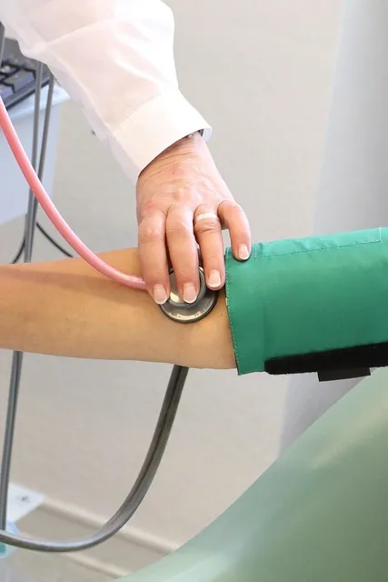
Current ATA guidelines recommend that levothyroxine-treated women with hypothyroidism should have their TSH checked at the first prenatal visit, with a target TSH of less than 2.5 mIU/L. If TSH is above this threshold, levothyroxine dose increase is recommended — typically a 25-30% increase as an initial empirical adjustment, often achieved by advising patients to take their usual daily dose plus two additional doses per week.
The recommended protocol for women with Hashimoto’s who are trying to conceive: achieve TSH below 2.5 mIU/L before conception. Once pregnancy is confirmed, immediately increase levothyroxine dose by approximately 25-30% and contact the prescribing physician for measurement of TSH within 4 weeks. Continue thyroid monitoring every 4-6 weeks throughout the first trimester and every trimester thereafter, or more frequently if doses are adjusted.
Women with Hashimoto’s who have normal thyroid function (euthyroid Hashimoto’s) and are not on thyroid hormone replacement present a different management question. Thyroid peroxidase antibodies (TPO-Ab) are associated with increased pregnancy loss risk even in euthyroid women — a meta-analysis found 2-3 times higher miscarriage rate in TPO-Ab positive euthyroid women compared to TPO-Ab negative women. Several small randomized trials have tested prophylactic low-dose levothyroxine in euthyroid TPO-Ab positive women, with conflicting results.
The TABLET trial (2019, NEJM) — the largest and most methodologically rigorous — found no benefit from low-dose levothyroxine in euthyroid thyroid antibody-positive women for preventing miscarriage or preterm birth. Other trials and meta-analyses have shown benefit, though. Current ATA guidelines do not recommend routine levothyroxine treatment for euthyroid TPO-Ab positive women, but acknowledge this remains a research area with ongoing uncertainty.
Hyperthyroidism in Pregnancy: Gestational Hyperthyroidism vs. Graves Disease
Low TSH in early pregnancy is common — but not all low TSH reflects pathological hyperthyroidism. Distinguishing gestational hyperthyroidism (transient, physiological, hCG-driven) from true Graves disease is clinically important, because management differs fundamentally.
Gestational hyperthyroidism occurs in approximately 1-3% of pregnancies and is driven by high hCG levels stimulating the thyroid. It typically presents in the first trimester, peaks with hCG levels at 10-12 weeks, and resolves spontaneously as hCG declines. It’s associated with hyperemesis gravidarum (severe pregnancy vomiting) — the same high hCG levels cause both thyroid stimulation and intractable nausea. It does not require antithyroid drug therapy and resolves without treatment.
Women with gestational hyperthyroidism do not have TSH-receptor antibodies (TRAb), distinguishing them from Graves disease.
Graves disease in pregnancy requires antithyroid drug treatment because maternal hyperthyroidism carries significant risks: miscarriage, preterm birth, maternal heart failure, and — importantly — transplacental passage of TSH-receptor-stimulating antibodies that can cause fetal and neonatal hyperthyroidism. The choice of antithyroid drug is critical and trimester-dependent.
Propylthiouracil (PTU) is preferred in the first trimester because methimazole is associated with a rare embryopathy — choanal atresia (blockage of nasal passages), aplasia cutis (scalp defects), and tracheoesophageal fistula — when used during organogenesis (first 10 weeks). PTU does not carry this teratogenic risk. After the first trimester, management should transition to methimazole, because PTU carries a risk of serious maternal hepatotoxicity (liver failure) with longer-term use.
This drug-switching protocol — PTU first trimester, methimazole second trimester onward — is established in guidelines but requires active coordination between the endocrinologist and obstetric team.
The management goal in Graves disease during pregnancy is to use the lowest possible antithyroid drug dose that maintains maternal free T4 in the upper normal or slightly above-normal range. The reasoning: antithyroid drugs cross the placenta and can cause fetal hypothyroidism if maternal doses are too high. Aiming for maternal free T4 in the slightly high-normal range ensures adequate fetal thyroid hormone while limiting fetal drug exposure — a more detailed target than standard hyperthyroidism management outside pregnancy.
Iodine Nutrition in Pregnancy: Getting It Right
Iodine requirements increase substantially during pregnancy — from approximately 150 mcg/day in non-pregnant adults to 220 mcg/day in pregnant women and 290 mcg/day in breastfeeding women. Iodine deficiency is the world’s most common preventable cause of intellectual disability. Worth sitting with that fact for a second, because it remains relevant even in developed countries where iodine sufficiency is assumed but not universal.
Mild-to-moderate iodine deficiency — below the threshold for overt hypothyroidism but insufficient for optimal thyroid hormone production during the heightened demands of pregnancy — is more common in developed countries than often recognized. A 2011 study in Lancet found that 66% of UK women had iodine intake below the recommended level during pregnancy.
US data from NHANES shows that while the US is generally iodine-sufficient at the population level, approximately 25-30% of pregnant women have urinary iodine concentrations suggesting insufficient intake.
Risk factors for iodine deficiency in pregnancy in developed countries include: avoiding iodized salt (common in people following low-sodium dietary patterns or using non-iodized salts); vegan or dairy-free diets (dairy products are the major iodine source in the US diet, not iodized salt); and use of non-iodine-containing prenatal vitamins (a 2014 survey found that fewer than 50% of US prenatal vitamins contain adequate iodine).
ATA guidelines recommend that pregnant and breastfeeding women in North America take a prenatal vitamin containing 150 mcg of potassium iodide daily, specifically to supplement dietary iodine. Kelp supplements are NOT recommended because of highly variable and potentially very high iodine content — excess iodine can paradoxically impair thyroid function in pregnancy through the Wolff-Chaikoff effect and exacerbate autoimmune thyroid disease. The recommendation is for measured potassium iodide supplementation in prenatal vitamins, not high-dose iodine preparations.
Postpartum Thyroiditis: The Condition Nobody Warns You About
The immunological basis of postpartum thyroiditis reflects the same immune dynamics that drive Graves disease remission during pregnancy and postpartum relapse. During pregnancy, immune tolerance is enhanced to protect the fetal semi-allograft. After delivery, this immune suppression lifts rapidly, creating an immunological rebound — a surge in autoimmune activity that can trigger thyroid inflammation in susceptible individuals.
Postpartum thyroiditis typically follows a biphasic pattern. The first phase (1-4 months postpartum) involves thyroid inflammation and cellular destruction, releasing stored thyroid hormones and causing transient hyperthyroidism with symptoms including palpitations, anxiety, insomnia, and heat intolerance. This phase is often misattributed to postpartum anxiety.
The second phase (4-8 months postpartum) involves the depleted thyroid struggling to produce adequate hormone, causing hypothyroidism with fatigue, depression, cold intolerance, weight gain, and brain fog — symptoms easily attributed to new-parent sleep deprivation rather than thyroid dysfunction.
Approximately 25-30% of postpartum thyroiditis cases result in permanent hypothyroidism, while the majority resolve within 12 months. Risk factors for permanent hypothyroidism include higher TPO-Ab titers and greater initial degree of hypothyroidism. Given this risk, women with significant postpartum thyroiditis should have thyroid function retested at 12 months postpartum and annually thereafter.
The treatment of postpartum thyroiditis is phase-dependent. The hypothyroid phase typically requires levothyroxine supplementation, though the dose can usually be reduced and discontinued as thyroid function recovers. The hyperthyroid phase does not respond to antithyroid drugs (the hyperthyroidism is from hormone release, not overproduction) and is managed with beta-blockers for symptom control if needed.
Distinguishing postpartum Graves disease — which CAN occur postpartum and is a distinct and more serious condition — from postpartum thyroiditis is clinically important and requires TSH-receptor antibody testing.
Monitoring Protocol for Thyroid Disease Throughout Pregnancy
A rational monitoring protocol for pregnant women with known thyroid disease, based on current evidence and ATA guidelines, looks substantially more intensive than monitoring outside pregnancy — appropriately so, given the physiological dynamics and the consequences of inadequate treatment.
For women on levothyroxine replacement before pregnancy: TSH testing at first prenatal visit (before 8 weeks ideally); immediate empiric dose increase of 25-30% or as directed by prescribing physician upon confirmed pregnancy; recheck TSH at 4-6 week intervals through the first trimester; monthly checks during second trimester if doses are stable; every 6 weeks in third trimester. After delivery, levothyroxine dose typically returns to the pre-pregnancy level immediately and TSH is checked at 6 weeks postpartum.
Women with Graves disease on antithyroid drugs should have TSH and free T4 checked every 4 weeks. TRAb (TSI) antibody levels should be checked at diagnosis, at 18-22 weeks, and near term to assess fetal hyperthyroidism risk — high maternal TRAb levels at 22-26 weeks predict neonatal Graves disease, allowing fetal surveillance to be intensified.
Neonatal surveillance after maternal Graves disease: TRAb crosses the placenta freely, and high maternal TRAb can cause neonatal Graves disease even when the mother is post-thyroidectomy or post-RAI (the antibodies persist even when the thyroid is gone). Neonatal thyroid function testing at 72 hours after birth, then at 1 week if maternal TRAb was elevated, is standard practice in high-risk neonates.
Reader Questions About Pregnancy Fundamentally Changes
Should I be tested for thyroid disease before trying to get pregnant?
Current ATA guidelines do not recommend universal thyroid screening before conception for the general population, but do recommend testing in women with specific risk factors: personal history of thyroid disease, thyroid surgery, or radioactive iodine treatment; family history of autoimmune thyroid disease; symptoms of thyroid dysfunction; presence of goiter; known thyroid antibodies; infertility or prior pregnancy loss; prior head/neck radiation; Type 1 diabetes or other autoimmune conditions.
Many fertility specialists routinely check TSH and TPO antibodies as part of pre-conception evaluation regardless of symptoms. If any of these risk factors apply, pre-conception thyroid evaluation is valuable and is the easiest time to optimize thyroid levels before pregnancy begins.
Can hypothyroidism cause miscarriage?
Yes — overt hypothyroidism (elevated TSH with low free T4) and subclinical hypothyroidism in the first trimester are consistently associated with increased miscarriage risk across multiple studies. The risk appears to be approximately 2-fold higher in women with elevated TSH compared to euthyroid women. TPO antibody positivity independently increases miscarriage risk even with normal TSH.
Whether treatment of subclinical hypothyroidism or thyroid antibody positivity reduces miscarriage rates is less certain — the TABLET trial showed no benefit from levothyroxine in antibody-positive euthyroid women, but treatment of frank hypothyroidism (elevated TSH) is generally recommended given the broader risk profile.
Is levothyroxine safe during pregnancy?
Yes — levothyroxine is one of the safest medications used in pregnancy. It is bioidentical to human T4 and has been used in pregnant women for decades without evidence of fetal harm when dosed appropriately to achieve normal thyroid hormone levels. Inadequate thyroid hormone replacement (under-treating hypothyroidism) is far more risky to the pregnancy and fetal development than levothyroxine therapy.
Concern about medication use in pregnancy is understandable. But forgoing necessary thyroid hormone replacement during pregnancy in the name of “avoiding medications” risks real harm to both maternal and fetal health.
My TSH is normal but I feel terrible — could thyroid be causing pregnancy symptoms?
Fatigue, cognitive fog, nausea, and mood changes are so common in normal pregnancy that distinguishing thyroid-related symptoms from normal pregnancy symptoms is genuinely difficult. Labs should guide management rather than symptoms alone in pregnancy.
However, remember that “normal” TSH requires using trimester-specific reference ranges — if first-trimester TSH is 3.5 mIU/L and the care team is using the standard non-pregnant upper limit of 4.0 mIU/L to call this normal, thyroid hormone may be inadequate for first-trimester demands in a woman with thyroid disease or antibodies. Requesting that TSH be interpreted using trimester-specific ranges and in the context of thyroid antibody status is appropriate advocacy.
What should I do about thyroid medication immediately after delivering?
Women who increased levothyroxine during pregnancy should generally return to their pre-pregnancy dose immediately after delivery, with TSH rechecked at 6 weeks postpartum. The increased demands of pregnancy resolve rapidly after delivery (hCG drops, TBG begins declining), and continuing the elevated pregnancy dose creates a risk of iatrogenic hyperthyroidism in the postpartum period.
The exception: some women with significant Hashimoto’s may need continued dose evaluation rather than automatic reduction, particularly if their pre-pregnancy dose was already at the lower end of adequacy. Postpartum thyroid function monitoring also screens for postpartum thyroiditis, which requires its own management approach distinct from pregnancy-related dose adjustments.
Thyroid Cancer Discovered During Pregnancy: Management Principles

The majority of thyroid cancers discovered during pregnancy are papillary thyroid microcarcinomas (PTC ≤1 cm) or low-risk papillary thyroid cancers — the same indolent tumors that dominate thyroid cancer incidence in the general population. The 2015 ATA thyroid cancer management guidelines specifically addressed pregnancy-associated thyroid cancer and provided guidance that has substantially reduced unnecessary surgery during pregnancy for these patients.
For low-risk papillary thyroid cancer discovered in the first or second trimester, active surveillance rather than surgery during pregnancy is now considered appropriate in most cases. Multiple prospective studies — primarily from Japan and Korea — demonstrate that properly selected low-risk PTC shows minimal growth during pregnancy and that deferring surgery until post-delivery does not worsen oncological outcomes.
A 2021 analysis of pregnancy-associated thyroid cancer outcomes found no significant difference in disease-free survival between patients treated surgically during pregnancy versus those who underwent post-partum surgery for equivalent-stage cancers.
If surgery is necessary — for rapidly growing tumors, tumors with lymph node involvement at diagnosis, or tumors causing significant compressive symptoms — the second trimester (weeks 14-26) is the safest operative window. First trimester surgery carries higher miscarriage risk from anesthesia and physiological stress; third trimester surgery increases risk of preterm labor. General anesthesia is generally safe in the second trimester for both mother and fetus when administered by experienced obstetric anesthesiologists.
Radioactive iodine treatment is absolutely contraindicated during pregnancy and breastfeeding — it is deferred until after delivery and cessation of breastfeeding.
TSH suppression — using high-dose levothyroxine to push TSH below normal as an adjunct therapy for thyroid cancer — is a strategy used after surgery in some thyroid cancer patients to reduce cancer cell stimulation by TSH. During pregnancy, TSH suppression must be balanced against the fetal risks of maternal hyperthyroidism.
The target TSH in thyroid cancer patients during pregnancy is typically 0.1-0.5 mIU/L (low but not fully suppressed) rather than the complete suppression sometimes used outside pregnancy — a compromise that reduces thyroid cancer stimulation while avoiding fetal thyroid dysfunction from maternal hyperthyroidism.
The Emotional Dimension of Thyroid Conditions in Pregnancy
Managing thyroid disease during pregnancy adds a layer of medical complexity to an already physically and emotionally demanding period. The psychological weight of navigating two simultaneous medical conditions — thyroid disease and pregnancy — while worrying about fetal wellbeing and the adequacy of treatment is not trivial. It deserves acknowledgment in clinical care that is often focused exclusively on physiological metrics.
Thyroid-related anxiety is common in pregnancy because the stakes feel higher. The knowledge that inadequate thyroid hormone during the first trimester can affect fetal brain development creates acute awareness of every symptom that might suggest suboptimal control. Pregnant women with Graves disease worry about TSI antibodies crossing the placenta and causing fetal hyperthyroidism. Women with Hashimoto’s worry that their TSH is high enough to matter.
This anxiety is rational — the risks are real — but it can become disproportionate to the statistical likelihood of harm when treatment is appropriate and monitoring is adequate.
Clear communication from care providers is the most effective intervention for pregnancy thyroid anxiety. Specifically: what the current lab values mean, what target values indicate adequate control, what the specific monitoring schedule is, and what symptoms should prompt immediate contact. This structure converts open-ended uncertainty (“my TSH might be wrong for pregnancy”) into manageable surveillance (“my next check is at 6 weeks, and I should call immediately if I develop palpitations or severe fatigue”).
The difference in anxiety load between these two experiences is substantial, and the clinical outcome is identical — but provider communication determines which experience the patient has.
Peer support from other women who have successfully navigated thyroid disease in pregnancy is often more emotionally sustaining than clinical information alone. Organizations like the American Thyroid Association, Thyroid UK, and dedicated online communities (carefully curated for evidence-based information) provide this peer dimension. The combination of accurate medical information and shared experience navigating similar challenges is more effective for reducing thyroid-related pregnancy anxiety than either alone.
Coordinated Care: Why Communication Between Providers Matters
Thyroid disease in pregnancy sits at the intersection of obstetrics and endocrinology — two specialties with different primary concerns, different reference ranges, different monitoring protocols, and occasionally different management philosophies. The coordination gap between these specialties is one of the most consistent sources of suboptimal care in this population, and patients who understand what effective coordination looks like can actively advocate for it.
The most common coordination failure: OB-GYNs using non-pregnancy TSH reference ranges to interpret first-trimester thyroid results, labeling values of 3-4 mIU/L as normal when trimester-specific ranges would classify them as above the recommended treatment threshold for thyroid antibody-positive patients. This occurs not from negligence but from the practical reality that many OB-GYNs manage the complete prenatal care of low-risk patients and may not be current on the subspecialty details of trimester-specific TSH management.
The solution isn’t to criticize OB-GYNs. It’s for patients to explicitly ask whether their TSH is being interpreted using pregnancy-specific reference ranges, and to request endocrinology co-management if thyroid disease management becomes complex.
Endocrinologists managing Graves disease during pregnancy need to communicate clearly with the obstetric team about: the antithyroid drug the patient is using and the trimester-specific drug preference, the target free T4 range appropriate for pregnancy-specific hyperthyroidism management, fetal surveillance recommendations based on TRAb levels, and the specific threshold for considering surgery during pregnancy versus post-partum management. This information should be documented in the shared medical record and communicated directly between providers, not just provided to the patient to relay.
High-risk maternal-fetal medicine (MFM) specialists should be involved when: TRAb levels are significantly elevated (predicting fetal/neonatal Graves risk requiring fetal surveillance), when surgical or radioactive iodine management decisions are being contemplated during pregnancy, when antithyroid drug side effects require dose changes that approach the boundary of fetal thyroid suppression risk, or when prior pregnancy losses suggest a complex thyroid-reproductive interaction. MFM specialists have both the fetal medicine expertise and the cross-specialty communication experience to coordinate complex thyroid-obstetric management effectively.
Patient-maintained personal health records — keeping copies of all thyroid labs, antibody values, medication doses, and ultrasound reports in a personal health record accessible at every appointment — dramatically improve coordination in fragmented healthcare systems. A patient who arrives at an obstetric appointment with a current thyroid record can immediately provide the context an OB-GYN needs to interpret a new TSH value accurately, rather than waiting for records to transfer between systems.
This kind of informed patient advocacy isn’t a burden. It’s a practical and empowering response to the real limitations of healthcare system coordination.
Long-Term Outcomes of Thyroid Disease in Pregnancy: What Research Shows
Understanding how thyroid disease in pregnancy affects both maternal and child outcomes provides the evidence base that makes appropriate thyroid management during pregnancy so important — and so worth the monitoring effort it requires.
Maternal outcomes of optimally treated thyroid disease during pregnancy are excellent. Women with Hashimoto’s hypothyroidism who achieve and maintain TSH below 2.5 mIU/L throughout pregnancy have pregnancy complication rates essentially comparable to euthyroid women without thyroid disease. The complications associated with poorly controlled hypothyroidism — gestational hypertension, placental abruption, preterm birth, increased cesarean rates — are substantially reduced or normalized with appropriate treatment. This is a situation where treatment genuinely changes outcomes rather than merely managing biomarkers.
Neurodevelopmental outcomes in children of mothers with treated thyroid disease have been tracked in multiple prospective cohorts. The CATS trial (Controlled Antenatal Thyroid Screening) in the UK followed children of mothers who received levothyroxine treatment versus standard care after universal TSH screening in early pregnancy. At 3 years, cognitive development was similar between groups — a finding initially interpreted as showing no benefit from screening and treatment.
However, the trial was criticized for starting treatment too late (median 13 weeks) to address the most critical period of fetal brain development (before 10 weeks) and for including women with very mild thyroid dysfunction. Follow-up at 9 years showed more complex results that remain under analysis. The CONTROL study (ongoing) is specifically designed to address earlier treatment timing and may provide more definitive data on neurodevelopmental benefit.
The long-term maternal autoimmune disease trajectory is also relevant: women with Hashimoto’s diagnosed during pregnancy have a higher lifetime risk of progression to overt hypothyroidism than the general population, and pregnancy itself accelerates this progression in some cases. Women who develop postpartum thyroiditis have a 25-50% probability of developing permanent hypothyroidism within 7 years.
Annual TSH monitoring in all women with documented postpartum thyroiditis is standard practice — and unfortunately, frequently overlooked in the absence of active symptoms, leaving new-onset hypothyroidism undetected for years. Consistent long-term follow-up is the final piece of comprehensive thyroid-pregnancy care.
The final consideration for long-term care is the well-documented relationship between autoimmune thyroid disease and other autoimmune conditions. Women with Hashimoto’s or a history of Graves disease have elevated lifetime risk for Type 1 diabetes, celiac disease, rheumatoid arthritis, lupus, and other autoimmune conditions — the same genetic HLA predispositions that allow one autoimmune condition also permit others.
This isn’t inevitability. It’s probability, and it argues for ongoing vigilance about new symptoms that could indicate emerging autoimmune disease rather than assuming any new symptom complex is “just” the thyroid. A woman who develops joint pain, fasting hyperglycemia, or recurrent GI symptoms after managing her thyroid disease for a decade deserves a fresh diagnostic evaluation — not a reflexive assumption that her thyroid medication needs adjustment.
The Practical Framework: Applying Pregnancy Fundamentally Changes Thyroid In Real Life
References