
Take a physician we’ll call Dr. Chen — a functional medicine doctor who’d been ordering hormone panels for twelve years. She understood, intellectually, that timing mattered for hormone testing. Understood it the way you understand a fact you’ve read a hundred times without it actually landing. Then a patient named David walked into her office with a testosterone result of 210 ng/dL, drawn at 3:45 PM after a stress-filled workday, utterly convinced he had severe hypogonadism. She retested him at 8 AM two days later. 612 ng/dL.
Same man. Same physiology. Numbers that told two completely different stories about his health.
Hormone testing is one of the most frequently misused tools in modern medicine and wellness practice. The numbers on a lab report look precise — to the decimal point, in some cases — and that precision creates a false sense of certainty. Hormones don’t sit still. They pulse, surge, cycle, respond to stress, respond to food, respond to light, respond to sleep, and respond to each other in feedback loops of extraordinary complexity. Nobody built them to be convenient to measure.
A hormone measurement is a snapshot of a system that never stops moving. The value of that snapshot depends entirely on when it’s taken, under what conditions, and how it gets interpreted against the clinical picture.
This piece is about optimizing hormone testing — the biological rhythms governing major hormone systems, the specific conditions that maximize diagnostic accuracy, and the testing strategies that separate signal from noise. Applies to testosterone, cortisol, thyroid hormones, estrogen and progesterone in women, insulin, growth hormone, and a handful of less commonly tested but clinically important biomarkers. Getting hormone testing right doesn’t just improve diagnostic accuracy. It can be the difference between appropriate treatment and years of mismanaged endocrine dysfunction.
The Circadian Foundation: Why Time of Day Changes Everything
The circadian clock — the roughly 24-hour biological rhythm synchronized mainly by light exposure — governs the secretion patterns of nearly every major hormone in the body. The master clock in the suprachiasmatic nucleus of the hypothalamus coordinates peripheral clocks in every organ and gland.
Test a hormone outside its peak secretion window and the result isn’t just low. It can be so far removed from that hormone’s normal functioning range that clinical interpretation becomes meaningless.
Testosterone follows one of the most pronounced circadian rhythms of any hormone in men. Levels peak early morning, typically between 6 and 10 AM, and decline steadily through the day. A study by Brambilla et al. published in the European Journal of Endocrinology followed testosterone levels in healthy men across a full 24-hour period and found morning levels 20-35% higher than afternoon levels, with some men showing intraday variation exceeding 50%.
The Endocrine Society guidelines explicitly recommend testosterone testing in men be performed between 7 and 10 AM, patient fasted overnight, adequately slept.
This circadian pattern is driven by the pulsatile release of LH (luteinizing hormone) from the pituitary, itself driven by pulsatile GnRH (gonadotropin-releasing hormone) from the hypothalamus. Both LH and GnRH show pronounced nighttime and early-morning surges. The testes respond to these LH pulses with testosterone production, which peaks a few hours after the morning LH surge. Testing at 3 PM, which is what happened to David the first time, captures the nadir of this rhythm.
Not wrong, exactly. It’s a real physiological state. But it’s not representative of peak androgenic function and shouldn’t be the basis for clinical decisions about testosterone deficiency.
The clinical implication is straightforward but widely ignored in primary care: a low testosterone result taken outside the morning window shouldn’t trigger a hypogonadism diagnosis without repeat morning testing. A 2017 audit published in JAMA Internal Medicine found a substantial minority of testosterone prescriptions in the United States were initiated based on a single test, often not collected under optimal conditions.
The downstream cost — unnecessary testosterone therapy, suppressed gonadal function from exogenous testosterone, potential long-term fertility compromise — is entirely avoidable with a simple protocol change.
Cortisol Testing: The Most Timing-Sensitive Hormone
If testosterone’s circadian variation is significant, cortisol’s is dramatic. The cortisol awakening response (CAR) — a surge of cortisol in the 30-45 minutes following waking — represents a 50-100% increase over basal levels. This isn’t just circadian rhythm. It’s a discrete physiological phenomenon driven by the anticipation of waking and early-morning light exposure, regulated by a different mechanism than the ongoing diurnal cortisol pattern.
Cortisol then declines from its morning peak through the afternoon, reaching its nadir around midnight to 2 AM.
Standard blood cortisol testing — a single serum draw — is almost worthless for evaluating adrenal function unless timing and conditions are precisely controlled. A single 8 AM serum cortisol has limited sensitivity for detecting subclinical adrenal insufficiency (Addison’s disease) because it only captures the peak, not the pattern. Tells you nothing about afternoon cortisol, evening cortisol, the robustness of the awakening response, or the slope of the daily decline.
Four-point salivary cortisol testing — collected at waking (immediately, before standing), 30 minutes after waking, early afternoon (noon to 2 PM), and evening (9-10 PM) — provides a far more diagnostically informative picture. Salivary cortisol measures the free, unbound fraction (the clinically active cortisol) rather than total cortisol including protein-bound fractions, and because saliva collection doesn’t require a clinic visit, samples can be collected under naturalistic conditions.
A 2012 systematic review in Psychoneuroendocrinology established that the cortisol awakening response measured in saliva is a reliable, valid marker of HPA axis reactivity, with established reference ranges.
The DUTCH complete hormonal test (Dried Urine Test for Comprehensive Hormones) extends this further by measuring cortisol metabolites — tetrahydrocortisol and tetrahydrocortisone — alongside free cortisol, offering insight into cortisol production rates and metabolic clearance. A person can have normal serum cortisol but elevated cortisol metabolites, suggesting the adrenals are producing more cortisol than usual while clearance keeps pace.
That’s a distinct clinical picture from someone with low metabolites and low free cortisol — which suggests genuinely reduced adrenal output.
Thyroid Testing: The Cascade That Demands Comprehensive Assessment
Thyroid hormone testing is a particularly fraught area, because standard care often relies on a single marker — TSH (thyroid-stimulating hormone) — which is both indirect and prone to timing artifacts. TSH follows a circadian rhythm, peaking between midnight and 4 AM with a nadir in the late afternoon. A study published in Thyroid found TSH values measured in the morning averaged 20-30% higher than afternoon values in the same patients.
In patients near the clinical cutoffs for hypothyroidism diagnosis (typically TSH above 4.0-4.5 mIU/L), afternoon testing can falsely normalize a morning-high TSH and delay diagnosis.
More fundamentally, TSH measures pituitary signaling, not thyroid hormone availability at the tissue level. Two patients with identical TSH values can have dramatically different free T4 and free T3 — the actual active thyroid hormones — because TSH reflects the pituitary’s assessment of thyroid hormone sufficiency, and the pituitary sees a slightly different hormonal environment than peripheral tissues do.
The clinical standard in functional medicine, and among a growing number of endocrinologists, is testing free T4, free T3, reverse T3 (rT3), and thyroid antibodies (anti-TPO, anti-TG) alongside TSH.
Free T3, the most biologically active thyroid hormone, is converted from T4 by deiodinase enzymes in peripheral tissues. The T4-to-T3 conversion can be impaired by numerous factors: selenium deficiency (deiodinase type I and II are selenoproteins), chronic inflammation (cytokines suppress DIO2 activity), caloric restriction, stress (cortisol promotes rT3 production instead of active T3), and certain medications including beta-blockers and amiodarone. A patient can have normal TSH and normal T4 while running inadequate T3 if conversion is impaired.
Testing T3 in isolation from T4 and rT3 misses this picture entirely.
Practical timing for thyroid testing: morning, fasting, before thyroid medication if applicable — taking thyroid medication before testing artificially elevates T4 and suppresses TSH. Patients on levothyroxine should take their morning dose after the blood draw. If afternoon testing is unavoidable, interpret TSH knowing it will trend lower than a morning value.
Test reverse T3 in the context of ongoing stress, illness, caloric restriction, or any clinical picture where T4 looks normal but hypothyroid symptoms persist anyway.
Female Hormone Testing: The Menstrual Cycle as the Testing Calendar

Test estradiol on day 21 without knowing the cycle length and confirming ovulation occurred, and the number that comes back is uninterpretable.
The standard protocol for reproductive endocrinology testing sets clear cycle-day windows for each marker. Baseline markers — FSH, LH, estradiol, AMH (anti-Müllerian hormone), and inhibin B — are tested on days 2-4 of the menstrual cycle (day 1 being the first day of full menstrual flow). This follicular-phase baseline captures ovarian reserve (FSH and AMH), pituitary function (LH), and baseline estrogenic activity before the follicular estradiol surge.
Progesterone is tested on day 21 of a standard 28-day cycle — or more precisely, 7 days after confirmed ovulation. In women with longer cycles, a 35-day cycle ovulates around day 21, so testing on cycle day 21 captures an early follicular sample, not a luteal-phase progesterone peak.
The approach favored by practitioners who pair cycle-tracking with hormone testing: use LH surge monitoring (urine OPK tests or digital fertility monitors) to pin down ovulation, then test progesterone exactly 7 days post-LH surge. A progesterone level above 10 ng/mL on that day confirms adequate luteal-phase progesterone production. Levels of 3-5 ng/mL may indicate anovulation or inadequate corpus luteum function even when the cycle looks regular on paper.
Testosterone in women peaks slightly in the pre-ovulatory phase (days 12-14) and stays relatively stable otherwise, which makes cycle timing less critical for testosterone than for progesterone or estradiol. SHBG, though — which shifts across the cycle in response to estrogen — affects free testosterone levels substantially. For clinical purposes, testing testosterone and SHBG in the early follicular phase (days 3-5) gives the most reproducible baseline.
Insulin and Blood Sugar: Fasting Is Not Optional
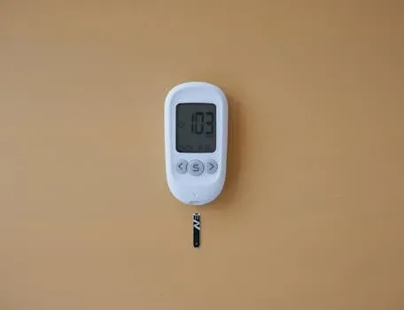
A fasting insulin level is the most sensitive early marker of insulin resistance available, and it requires a true twelve-hour overnight fast — no calorie intake, no coffee with cream, ideally no intense exercise in the preceding 24 hours (which can acutely lower insulin sensitivity and produce a misleadingly favorable reading).
A fasting insulin above 10 μIU/mL is broadly considered a signal of insulin resistance even when fasting glucose looks completely normal. Research by Kraft (2008), using five-hour oral glucose tolerance tests with insulin assays at multiple time points, showed that the vast majority of people who will eventually develop type 2 diabetes display hyperinsulinemic patterns in response to a glucose challenge for years before fasting glucose abnormalities ever show up.
Fasting insulin is the least sensitive part of this picture. It’s also the most practical to test in a normal clinical setting.
The two-hour postprandial insulin test — measured two hours after a standard 75g glucose challenge — gives substantially more information than fasting insulin alone. HOMA-IR (homeostatic model assessment of insulin resistance), calculated as fasting insulin × fasting glucose / 405 (glucose in mg/dL), is a validated surrogate for insulin resistance built from readily available markers. HOMA-IR above 1.9 is associated with insulin resistance; above 2.9 is a strong signal.
The calculation requires simultaneous fasting glucose and insulin, both collected after a proper overnight fast.
Growth Hormone: The Pulse Problem
Growth hormone is maybe the most extreme example of the timing problem in hormone testing. GH gets secreted in discrete pulses, the largest occurring during deep sleep — specifically the first slow-wave sleep cycle, typically 60-90 minutes after sleep onset. Between pulses, GH levels fall to essentially zero.
A random serum GH test drawn at any point during waking hours is nearly guaranteed to show near-zero levels in a normal adult. Completely useless for assessing GH secretory capacity.
The practical workaround is IGF-1 (insulin-like growth factor 1), produced in the liver in response to GH stimulation, with a half-life of roughly 15-20 hours — making it a stable, reproducible marker of integrated GH secretion over time. IGF-1 testing doesn’t require specific timing, though a consistent morning fasting draw is standard for reproducibility.
The limitation: IGF-1 can be low for non-GH reasons — malnutrition, liver disease, insulin deficiency (IGF-1 production requires insulin co-stimulation), genetic variation in the GH receptor. Elevated IGF-1 despite normal GH secretion is also possible in states of insulin excess.
For formal GH deficiency assessment, provocative testing is required — insulin tolerance test (ITT), GHRH-arginine test, or glucagon stimulation test. These deliberately provoke a GH pulse by manipulating the regulatory inputs (hypoglycemia from insulin stimulation, or direct GHRH plus arginine stimulation) and measuring the peak response. Adult GH deficiency is defined as a GH peak below 11 ng/mL on the ITT or below 9 ng/mL on the GHRH-arginine test (cutoffs vary slightly by assay and guidelines).
DHEA-S, Pregnenolone, and the Adrenal Reserve Markers
DHEA-S (dehydroepiandrosterone sulfate) has a relatively flat diurnal variation compared to cortisol — no dramatic morning-peak-to-evening-trough pattern — which makes it one of the more timing-tolerant adrenal markers. That said, DHEA-S gets acutely suppressed by illness, caloric restriction, chronic psychological stress, and by the same cortisol-dominant states that define HPA axis dysfunction.
Test it fasted (food can modestly alter DHEA-S) during a period of relative health — not during acute illness, not immediately after major physiological stress.
Pregnenolone — the “mother hormone” upstream of both cortisol and DHEA — is less commonly tested but offers real insight into adrenal substrate availability. Low pregnenolone can limit production of downstream hormones including DHEA, progesterone, testosterone, and cortisol. Pregnenolone testing is most informative in a fasting morning draw. Reference ranges are age-dependent and decline substantially with age, but clinical labs often use ranges based on young adults, which can mask meaningful declines in older populations.
The “cortisol steal” hypothesis — the idea that chronic stress shunts pregnenolone preferentially toward cortisol production, reducing DHEA and sex hormone output — is mechanistically plausible but not definitively established in human research. The concept has been used to market adrenal support supplements. Sometimes aggressively.
What’s well-established: chronic HPA axis activation does suppress DHEA-S, low DHEA-S correlates with adverse health outcomes including cardiovascular disease and all-cause mortality across multiple longitudinal studies, and restoring DHEA-S to youthful levels has shown some benefit in clinical trials for fatigue, libido, bone density, and immune function. The interpretation should be clinical, not purely biochemical.
Testing Estradiol in Men: The Assay Problem
Men testing estradiol run into a specific technical problem: most standard estradiol assays are built and calibrated for women, whose baseline estradiol runs 50-400 pg/mL across the menstrual cycle. Male estradiol levels typically sit at 10-40 pg/mL. Standard assays operating at the bottom of their detection range in male samples produce unreliable results — a problem that’s caused real confusion in clinical practice.
The fix is the “sensitive estradiol” or “ultrasensitive estradiol” assay, which uses liquid chromatography-mass spectrometry (LC-MS/MS) instead of immunoassay. LC-MS/MS delivers accurate estradiol quantification down to 1-2 pg/mL — fully appropriate for the male reference range. When ordering estradiol for men, or for postmenopausal women, specifically request the LC-MS/MS assay. In the United States, LabCorp offers “Estradiol, Ultrasensitive, LC/MS/MS” (test #140244) and Quest offers “Estradiol, Ultrasensitive” (test #30289).
Generic “estradiol” orders default to immunoassay. Not good enough.
For male estradiol timing: unlike testosterone, estradiol doesn’t show a strong circadian variation in men. A morning draw is still preferable for reproducibility and to correlate with morning testosterone testing, but an afternoon draw won’t badly misrepresent male estradiol the way it misrepresents testosterone. Calculate the testosterone:estradiol ratio from the same blood draw to eliminate timing artifacts between the two values.
Practical Timing Protocol: A Specimen Collection Guide
Optimizing hormone testing means standardizing collection conditions — not just time of day, but the physiological state at collection. What follows is evidence-based best practice for a comprehensive male hormone panel. For women, the cycle-day considerations above apply on top of these conditions.
Timing: 7 AM to 9 AM is the target window for maximum diagnostic utility. Captures peak testosterone, the declining phase of the cortisol awakening response (which can be folded into the collection), and early-morning thyroid patterns. Fasting: overnight, 10-12 hours, water only. Exceptions: thyroid medications should be taken after the draw; other medications should be discussed with a clinician regarding hold periods.
Sleep: the preceding night should be representative — not a recovery night after severe deprivation, not an unusually heavy sleep either. Minimum six hours required; seven to eight is ideal.
Exercise: avoid intense exercise in the 24-48 hours before testing. Acute exercise transiently elevates testosterone (for 15-30 minutes post-workout), elevates cortisol substantially and persistently for hours, and can alter thyroid hormone patterns. The blood draw itself is a mild stressor that transiently elevates cortisol — minimized when the patient has sat quietly for 15-20 minutes beforehand. Some labs offer “resting” specimen collection protocols for cortisol-sensitive testing.
Alcohol: avoid for 48-72 hours before testing. Alcohol suppresses testosterone through multiple mechanisms (direct Leydig cell toxicity, elevated NADH ratio impairing testosterone synthesis, elevated cortisol, altered LH pulsatility) and elevates estradiol by inducing liver alcohol dehydrogenase pathways that interact with estrogen metabolism. Even moderate social drinking the night before can shift morning testosterone meaningfully lower.
Interpreting Patterns vs. Single Values
The most sophisticated approach to hormone testing isn’t optimizing the conditions for a single time-point measurement. It’s accepting that endocrine function is inherently dynamic and building a testing strategy that captures patterns over time. A single well-collected morning testosterone tells you something. Three morning testosterone values collected over six weeks, under consistent conditions, tells you considerably more. The coefficient of variation for serum testosterone in healthy men — tested under standardized conditions on different days — runs approximately 10-15%.
Which means a value of 500 ng/dL could legitimately test anywhere from 425 to 575 ng/dL on a given day, same person.
Hormone patterns that should trigger concern and repeat testing: any single value outside the reference range (repeat before acting); any significant divergence between correlated hormones (normal testosterone with elevated LH suggests testicular resistance; low testosterone with low LH suggests central hypogonadism — the pattern directs the diagnosis); and any combination of clinical symptoms with borderline lab values. Symptoms matter. A man with significant low-testosterone symptoms and a testosterone of 320 ng/dL deserves more diagnostic attention than a man with identical lab values and no symptoms at all.
Trend testing — retesting at 6-12 month intervals — reveals the trajectory of hormonal change that single snapshots completely miss. A testosterone that has declined from 720 to 480 ng/dL over three years, while still “normal,” is a clinically important finding that warrants investigation and intervention. The current lab value sits within reference range. The trend does not — expected testosterone decline is approximately 1-2% per year in men, and this isn’t that.
Catching and reversing that trajectory while values are still normal is fundamentally better medicine than waiting for a value to cross below the reference range cutoff and reacting only then.
Circadian Foundation Time Q&A
My testosterone was tested at 2 PM and was 350 ng/dL. Should I start TRT?
No — not based on that test alone. An afternoon testosterone result is not a valid basis for diagnosing hypogonadism or initiating testosterone replacement therapy. The Endocrine Society guidelines require two morning fasting tests showing low testosterone (typically below 300 ng/dL) before initiating TRT, along with clinical symptoms. Get retested at 7-9 AM, fasting, after adequate sleep, and without recent intense exercise.
Many men who present with 2 PM testosterone values in the 300-400 ng/dL range test 500-650 ng/dL under proper conditions. Proper timing can literally save a man from unnecessary lifelong hormone replacement therapy.
Do I need to fast for thyroid testing?
Technically, TSH and thyroid hormone levels are minimally affected by fasting status in most people. Fasting is still recommended for standardization, though — particularly if testing alongside metabolic markers like insulin and glucose, and to eliminate the confounding effect of food-induced changes in cortisol, insulin, and other hormones that interact with thyroid signaling. The more critical variable for thyroid testing is morning timing, consistency across serial tests, and making sure thyroid medication wasn’t taken before the draw.
If someone on levothyroxine takes it before the blood draw, T4 will appear artificially elevated and TSH artificially suppressed — a misleading clinical picture.
Can stress before the blood draw affect my hormone results?
Yes, substantially — particularly for cortisol and testosterone. Acute psychological stress before and during a blood draw activates the HPA axis, elevating cortisol within minutes. Cortisol suppresses LH and testosterone acutely, meaning the stress of the clinic visit itself — traffic, rushing, anxiety about needles — can lower testosterone by 10-20% at the moment of the draw. Standard recommendation: sit quietly for 15-20 minutes before venipuncture.
Some practitioners recommend lying down during cortisol collection for maximum standardization. Significant acute stress before the draw — a frightening event, major conflict, panic — warrants rescheduling.
Is the DUTCH test better than blood testing for hormones?
It’s not a replacement for blood testing. It’s a complement. The DUTCH test measures urinary hormone metabolites, which provides information about metabolic pathways — how estrogen is being processed, how cortisol production and clearance rates compare — that serum testing doesn’t capture. It also provides multi-point cortisol data across the day.
What it doesn’t provide is the real-time serum levels that standard clinical reference ranges are built on — so for diagnosing testosterone deficiency or thyroid disease, serum testing remains the clinical standard. DUTCH is particularly valuable for assessing cortisol patterns, estrogen metabolism, and melatonin, and for functional medicine applications where understanding metabolic pathways matters as much as absolute levels.
How often should I retest hormone levels?
For a healthy adult doing periodic wellness monitoring without active treatment: annually is reasonable, always collected under the same standardized conditions to keep year-over-year comparison valid. For someone actively managing a hormonal condition with supplements, lifestyle changes, or medication: every eight to twelve weeks during the optimization phase, then quarterly once stable. For TRT patients: every three months for the first year (monitoring hematocrit, PSA, testosterone and estradiol levels), then every six months once stable.
Serial testing under consistent conditions is what makes the data useful. Comparing a 9 AM fasting testosterone from last January with a 3 PM non-fasting testosterone from this September tells you nothing meaningful. Consistency of collection conditions isn’t optional. It’s the entire basis of hormonal trend monitoring.
Age-Specific Considerations in Hormone Testing Timing
Hormonal rhythms change with age in ways that affect optimal testing windows. In adolescent males, the testosterone circadian rhythm is more pronounced and the morning peak sharper, making the 7-9 AM window even more critical. In men over sixty, the circadian amplitude of testosterone declines — morning-to-afternoon variation shrinks to around 15-20% rather than 30-50% — but morning testing remains the standard regardless.
In postmenopausal women, the dramatic cycle-dependent hormone variation disappears, and testing can happen at any time, though morning fasting draws remain preferred for the metabolic markers tested alongside reproductive hormones.
In children suspected of growth hormone deficiency, single random GH measurements are unreliable at any time because of episodic secretion, but IGF-1 testing should still happen in a morning fasting state, with the caveat that IGF-1 is normally lower in children with delayed puberty, severely underweight children, and during illness. Pediatric endocrinology reference ranges are age- and Tanner-stage stratified — apply adult reference ranges to children and the conclusions come out systematically wrong.
For women in perimenopause — typically the five to ten years before the final menstrual period — cycle length becomes irregular, and FSH levels start rising erratically. FSH testing on cycle day 2-3 can show dramatically different values month to month during perimenopause, because the underlying ovarian reserve is declining and the hormonal feedback loops are destabilizing. Serial FSH measurement across multiple cycles is more informative than any single result.
AMH (anti-Müllerian hormone), which has less day-to-day variation than FSH, gives a more stable read on remaining ovarian follicle pool and can be tested at any point in the cycle — a valuable anchor measurement in perimenopausal assessment.
In men over fifty, the morning testosterone advantage is less pronounced, but the other conditions — fasting, adequate prior sleep, absence of acute stress, absence of alcohol — matter just as much as ever.
Older men are more likely to be on medications that affect hormone levels: statins modestly reduce testosterone by affecting cholesterol availability (testosterone is synthesized from cholesterol); opioids dramatically suppress the HPG axis (opioid-induced hypogonadism is a clinically underappreciated consequence of chronic opioid use); glucocorticoids suppress testosterone, DHEA, and thyroid-stimulating signaling. Any comprehensive hormone panel in an older adult needs a careful medication review as part of interpretation.
David, from the opening, eventually got his proper 8 AM testosterone result. He wasn’t hypogonadal. He was a man whose demanding job had put his doctor’s appointment — and his blood draw — at the end of a cortisol-saturated afternoon. The testosterone his body produced was fine. The conditions under which it got measured were not. That distinction, between what the body produces and what a measurement captures, is the entire point of optimizing hormone testing timing.
The laboratory value and the biological reality are only the same thing when the collection conditions honor the rhythms of the system being measured.
For most hormones, optimizing testing timing doesn’t require extraordinary effort: morning draws before 9 AM, overnight fast, adequate prior sleep, no recent exercise, no alcohol. Not burdensome constraints. They’re simply the conditions under which the measurement means what clinicians assume it means. Ignoring them doesn’t save time. It generates data that either misleads or has to be repeated. Getting it right the first time, under standardized conditions, is the foundation everything else in endocrine assessment gets built on.
The Hidden Cost of Inconsistent Testing Conditions
Consider the systemic problem: millions of hormone tests get ordered every year in primary care settings where the ordering physician has no specific training in endocrinology, the lab staff has no protocol for standardizing collection conditions, and the patient has no idea that showing up after a large breakfast, a stressful commute, and a cup of coffee with cream just rendered the result clinically questionable.
The result gets reported, compared to a reference range, and a clinical decision gets made. Possibly the wrong one.
A 2019 analysis of testosterone testing patterns in a large US health system found nearly 40% of testosterone tests were collected after 10 AM, and less than half of men with an initial low result were retested before treatment decisions were made. Not edge cases. That’s the majority of clinical hormone testing happening in the real world. The problem isn’t the tests. It’s the unstandardized, context-insensitive way they’re deployed.
The fix requires education at two levels: the ordering clinician, who should specify collection conditions on the order and follow up with repeat testing before acting on a single result; and the patient, who should understand why these conditions matter and advocate for proper testing.
The latter matters more every year as direct-to-consumer hormone testing proliferates — services that mail test kits without providing guidance on collection conditions create the worst possible scenario: lab-grade analytical precision applied to samples collected under arbitrary conditions.
Hormone testing, done right, is one of the most powerful diagnostic tools in modern medicine. It can catch thyroid dysfunction before symptoms become debilitating, catch adrenal insufficiency before an adrenal crisis, identify insulin resistance years before diabetes develops, and characterize reproductive hormone deficiencies that explain years of unexplained fatigue, depression, and sexual dysfunction. None of that power is accessible if the testing conditions aren’t controlled.
The hormone system is beautiful in its complexity, responsive in its dynamics. Those dynamics are only visible when measured correctly.
The circadian system evolved over hundreds of millions of years to create rhythmic predictability in biological processes. Hormones peak when needed, decline when not, and create a precisely timed cascade of physiological events across the 24-hour day. Measure these hormones and the target is, by definition, moving.
The art of hormone testing optimization is choosing the right moment in the rhythm — and the right conditions around that moment — so the sample actually represents the biological reality it’s supposed to capture. Everything else is just numbers on a page.
The Practical Framework: Applying Circadian Foundation Time Day In Real Life
References