
Maria noticed the lump while applying sunscreen before a beach vacation. She almost dismissed it — maybe a swollen lymph node from the cold she’d had two weeks ago. Her husband noticed her pressing her fingers to her neck again at dinner, and something in his expression made her call her doctor the next morning. That single phone call, made on impulse, changed the trajectory of what came next.
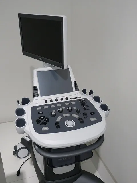
Her ultrasound flagged a 1.4-centimeter nodule with irregular borders and microcalcifications. Fine-needle aspiration biopsy confirmed papillary thyroid carcinoma. Maria was 38, otherwise healthy, and completely blindsided. She had no family history, never smoked, ate reasonably well. The diagnosis landed like something from a different person’s life.
Thyroid cancer is among the most common endocrine malignancies diagnosed today, and its incidence has tripled over the past three decades — though researchers debate how much of that reflects genuine increases versus improved detection technology. Understanding what thyroid cancer actually is, how to think about risk and surveillance, and what the evidence says about treatment outcomes is essential for anyone navigating this diagnosis or supporting someone who is.
THE ANATOMY OF THE THYROID AND WHY CANCER ARISES THERE
The thyroid gland sits at the base of the throat, shaped roughly like a butterfly with two lobes connected by a narrow isthmus. It weighs between 20 and 30 grams in most adults — less than an ounce — yet exerts metabolic influence over virtually every cell in the body. The gland produces thyroxine (T4) and triiodothyronine (T3), hormones that regulate energy metabolism, body temperature, heart rate, protein synthesis, and the sensitivity of other hormone systems.
Thyroid tissue is composed primarily of follicular cells, which produce T4 and T3, and parafollicular C cells, which produce calcitonin. Cancer can arise from either cell type, producing distinct tumor subtypes with very different biological behaviors and prognoses. The follicular cells give rise to the most common thyroid cancers — papillary and follicular — while C cells produce medullary thyroid carcinoma.
Carcinogenesis in thyroid tissue follows the same general rules as cancer elsewhere: genetic mutations accumulate in ways that allow cells to evade normal growth controls, acquire the ability to invade local tissue, and sometimes spread to distant sites. In papillary thyroid carcinoma, the most common subtype, the BRAF V600E mutation is present in roughly 60 percent of cases. This mutation activates the MAPK/ERK signaling pathway, driving uncontrolled cellular proliferation.
RET/PTC gene rearrangements, which were identified as significant risk factors following studies of Chernobyl-exposed populations, are found in another 20-30 percent of papillary cases.
Radiation exposure remains the most clearly established environmental risk factor. Childhood exposure to ionizing radiation — whether from medical treatments, nuclear fallout, or occupational sources — significantly increases lifetime thyroid cancer risk. The thyroid gland is particularly sensitive to radiation damage during childhood because of its rapid cellular turnover and active iodine uptake.
Studies of populations exposed to radioactive iodine from the Chernobyl disaster showed a dramatic increase in papillary thyroid carcinoma among those who were children at the time of exposure, with latency periods of 5-10 years.
THE FOUR MAIN TYPES AND HOW THEY DIFFER
Thyroid cancer is not a single disease. The four main histological subtypes have dramatically different biological behaviors, treatment responses, and outcomes. Grouping them under one umbrella obscures critical distinctions that matter enormously to patients.
Papillary thyroid carcinoma accounts for approximately 80-85 percent of all thyroid cancers. It grows slowly, tends to spread first to regional lymph nodes rather than distant organs, and carries an excellent prognosis — 10-year survival rates exceed 95 percent for localized disease. The distinctive “Orphan Annie eye” nuclear features seen on pathology reflect the characteristic empty-appearing nuclei that define this subtype. Even with lymph node involvement, outcomes remain favorable.
The challenge with papillary carcinoma is not the biology but the decision-making around how aggressively to treat what is often an indolent disease.
Follicular thyroid carcinoma represents 10-15 percent of cases. Unlike papillary carcinoma, it tends to spread via the bloodstream to distant sites — lungs and bone most commonly — rather than through lymphatic routes. Distinguishing follicular carcinoma from follicular adenoma (a benign tumor) requires surgical excision because the defining feature, capsular or vascular invasion, cannot be assessed by biopsy alone.
This creates a clinical dilemma: many patients with follicular neoplasms diagnosed on fine-needle aspiration ultimately have benign disease but require surgery to be certain.
Medullary thyroid carcinoma arises from parafollicular C cells and constitutes roughly 3-4 percent of thyroid cancers. About 25 percent of medullary cases are hereditary, associated with mutations in the RET proto-oncogene and occurring in the context of Multiple Endocrine Neoplasia type 2 (MEN2). Genetic testing is therefore essential for every patient diagnosed with medullary carcinoma. It does not take up iodine, making radioactive iodine ineffective.
Prognosis depends heavily on stage at diagnosis; localized disease carries good outcomes but regional or distant metastases significantly worsen survival.
Anaplastic thyroid carcinoma is rare — under 2 percent of cases — but extremely aggressive. It typically presents in older adults, grows rapidly, and is nearly uniformly fatal within months. The median survival after diagnosis is approximately 5 months. Unlike the other subtypes, anaplastic carcinoma often derives from dedifferentiation of a pre-existing differentiated thyroid cancer, which is one reason aggressive surveillance and treatment of known thyroid cancer matters even when the initial tumor appears indolent.
THE INCIDENCE DEBATE: GENUINE EPIDEMIC OR DETECTION ARTIFACT?
Thyroid cancer incidence has tripled in the United States since the 1970s, a trend mirrored across many high-income countries. The question of whether this represents a genuine increase in cancer burden or a detection artifact has significant implications for how aggressively clinicians should pursue thyroid nodules found incidentally.
A landmark 2014 analysis by Louise Davies and Gilbert Welch in JAMA Internal Medicine examined thyroid cancer incidence and mortality data from 1975 to 2009. They found that while incidence had tripled, mortality had remained essentially flat — increasing only 0.19 percent annually. If the new cancers being detected were genuinely lethal, mortality would have risen proportionally.
The flat mortality curve strongly suggests that much of the increased incidence reflects detection of subclinical cancers that would never have caused symptoms or death — a phenomenon called overdiagnosis.
South Korea provides a natural experiment in this regard. After a government-sponsored ultrasound screening program was launched in the late 1990s, thyroid cancer incidence increased 15-fold between 1993 and 2011. Following publication of research highlighting this pattern, screening rates dropped dramatically. Thyroid cancer incidence subsequently fell, without any corresponding increase in mortality. This provides compelling evidence that a substantial proportion of detected thyroid cancers, particularly small papillary microcarcinomas, represent overdiagnosed disease.
The clinical and ethical implications are significant. A cancer diagnosis carries psychological weight, treatment risks, and long-term surveillance burdens. If many detected thyroid cancers would never cause harm, the net benefit of aggressive detection and treatment becomes uncertain. This has driven a movement toward active surveillance protocols for low-risk papillary microcarcinomas, pioneered by Akira Miyauchi and colleagues at Kuma Hospital in Japan.
RISK FACTORS BEYOND RADIATION: WHAT THE EVIDENCE SHOWS
Iodine status occupies a complex position. Both iodine deficiency and iodine excess have been associated with different thyroid cancer subtypes. Iodine deficiency historically correlated with higher rates of follicular thyroid carcinoma, while regions with high iodine intake show higher rates of papillary carcinoma. This may reflect differential effects on thyroid-stimulating hormone dynamics and cellular proliferation patterns. The introduction of iodized salt in many countries coincided with a shift in the histological distribution of thyroid cancers toward the papillary subtype.
Obesity and insulin resistance have emerged as potential modifiable risk factors. Several epidemiological studies have found associations between higher body mass index and thyroid cancer risk, with some evidence suggesting that insulin and insulin-like growth factor 1 (IGF-1) signaling may stimulate thyroid cell proliferation. A 2021 meta-analysis in Obesity Reviews found a statistically significant positive association between obesity and thyroid cancer risk, with a relative risk of approximately 1.55 for obese individuals compared to normal-weight controls.
Thyroid-stimulating hormone (TSH) levels within the normal range may also influence risk. Higher TSH levels, even within the reference range, have been associated with greater likelihood of malignancy in thyroid nodules. TSH is a potent growth factor for thyroid tissue, and chronic mild elevation — as occurs in subclinical hypothyroidism or in states of relative iodine deficiency — may promote cellular proliferation that increases cancer risk over time.
Family history confers meaningful risk. First-degree relatives of papillary thyroid carcinoma patients have a 4-10-fold higher risk of developing the disease compared to the general population. The genetic basis for familial non-medullary thyroid cancer is not fully understood; unlike medullary carcinoma, no single dominant mutation accounts for most familial clustering. Multiple susceptibility loci identified through genome-wide association studies each contribute modest risk increases.
DIAGNOSIS: NODULES, BIOPSY, AND THE TIRADS SYSTEM
Most thyroid cancers are found not because of symptoms but because of thyroid nodules detected incidentally during imaging performed for other purposes — a neck CT for trauma, a carotid ultrasound for cardiovascular evaluation, an MRI for cervical spine pain. This incidental discovery pathway creates immediate decisions about how to evaluate and whether to biopsy.
Thyroid nodules are extremely common. Ultrasound detects palpable nodules in 4-7 percent of adults, but high-resolution ultrasound reveals nodules in 19-67 percent of randomly selected populations. The vast majority are benign — cysts, colloid nodules, hyperplastic nodules, follicular adenomas. The challenge is distinguishing the small minority that harbor malignancy without subjecting everyone to invasive procedures.
The Thyroid Imaging Reporting and Data System (TIRADS) provides a standardized framework for this risk stratification. Developed by the American College of Radiology and published in 2017, TIRADS assigns risk scores based on ultrasound features: composition (solid versus cystic), echogenicity, shape, margins, and echogenic foci. Each feature contributes points; higher total scores indicate greater malignancy risk and guide decisions about biopsy thresholds.
The system also incorporates nodule size, because the clinical significance of a 2 cm versus 0.4 cm nodule with identical sonographic features differs substantially.
Fine-needle aspiration biopsy (FNA) remains the gold standard for tissue diagnosis. A thin needle is inserted into the nodule under ultrasound guidance, and cells are aspirated for cytological analysis. Results are reported using the Bethesda System, a six-tier classification from non-diagnostic (Category I) through malignant (Category VI). Bethesda Category III (atypia of undetermined significance) and Category IV (follicular neoplasm) represent the diagnostic gray zones where molecular testing has significantly improved clinical decision-making.
Molecular tests such as Afirma Gene Sequence Classifier and ThyroSeq v3 analyze genetic material from FNA samples to refine malignancy risk estimates for indeterminate nodules. Afirma uses a 167-gene expression classifier; a “benign” result reduces the false-negative rate enough that many clinicians feel comfortable avoiding diagnostic surgery for appropriately selected patients. ThyroSeq v3 detects mutations, gene fusions, and copy number variants across 112 genes. These tests have meaningfully reduced unnecessary thyroid surgeries for indeterminate nodules.
SURGICAL TREATMENT: TOTAL THYROIDECTOMY VERSUS LOBECTOMY

The 2015 American Thyroid Association guidelines represented a significant shift toward more conservative surgery for appropriately selected patients. For unifocal papillary thyroid microcarcinomas (tumors 1 cm or smaller) without high-risk features, active surveillance without immediate surgery became an acceptable management strategy. For papillary cancers between 1 and 4 cm without extrathyroidal extension, aggressive lymph node involvement, or distant metastases, lobectomy was recognized as a reasonable alternative to total thyroidectomy.
The operative risks of thyroid surgery, while generally low in experienced hands, are not trivial. Total thyroidectomy carries risks of permanent hypoparathyroidism (damage to the parathyroid glands, causing chronic low calcium levels) and recurrent laryngeal nerve injury (causing vocal cord paralysis and voice changes). These complications occur in roughly 1-2 percent of cases even at high-volume centers, and at higher rates in less experienced surgical settings. Lobectomy substantially reduces these risks by halving the scope of dissection.
The trade-off of lobectomy is that patients may retain functioning thyroid tissue and potentially avoid lifelong thyroid hormone replacement — though approximately 30-45 percent of lobectomy patients eventually require supplementation. The retained lobe must be monitored for recurrence. And if subsequent pathology reveals features that would have indicated total thyroidectomy, completion surgery is necessary, carrying all the risks of reoperative neck surgery.
Surgeon volume matters enormously. Studies consistently demonstrate that high-volume thyroid surgeons — those performing more than 100 thyroid surgeries annually — achieve significantly lower complication rates than low-volume surgeons. A 2013 study in Annals of Surgery found that high-volume surgeons had permanent hypoparathyroidism rates of 0.5 percent versus 3.8 percent for low-volume surgeons. Seeking care at a center with thyroid surgical volume matters more than almost any other treatment decision for most patients.
RADIOACTIVE IODINE: WHEN IT HELPS AND WHEN IT DOESN’T
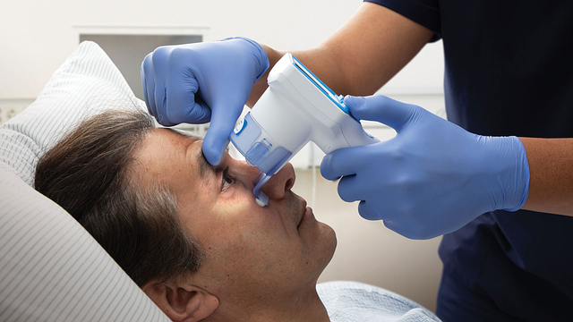 Radioactive iodine (RAI, I-131) therapy exploits the unique ability of differentiated thyroid tissue to concentrate iodine. After total thyroidectomy removes most thyroid tissue, any remaining thyroid cells — including metastatic differentiated thyroid cancer cells — continue to express the sodium-iodide symporter and will avidly take up administered radioiodine. The radiation then destroys those cells from within, achieving ablation without significant exposure to surrounding tissues.
For high-risk differentiated thyroid cancers — large tumors with extrathyroidal extension, extensive lymph node involvement, or distant metastases — RAI adjuvant therapy after total thyroidectomy reduces recurrence risk and improves survival. This benefit is not contested. The controversy concerns intermediate and low-risk cancers, where evidence for benefit is much weaker and historical use has been described as excessive.
A pivotal 2012 randomized controlled trial published in the New England Journal of Medicine (the ESTIMABL trial) compared different RAI doses and preparation strategies in low-risk patients. It found that low-dose RAI (1.1 GBq) was non-inferior to high-dose (3.7 GBq) for thyroid remnant ablation in this population, and that thyroid hormone withdrawal could be replaced by recombinant human TSH (rhTSH) stimulation for RAI preparation, avoiding the miserable hypothyroid state previously required.
Subsequent research has challenged whether low-risk patients benefit from RAI at all, and current guidelines discourage routine RAI use for low-risk differentiated thyroid cancers.
Preparation for RAI requires TSH elevation above 30 mIU/L to maximize iodine uptake. This can be achieved by withdrawing thyroid hormone for 4-6 weeks (causing clinical hypothyroidism with fatigue, cognitive slowing, depression, weight gain, and fluid retention) or by administering rhTSH injections (Thyrogen) while continuing thyroid hormone replacement. Where available and insurance permits, rhTSH preparation is strongly preferred for patient quality of life.
TSH SUPPRESSION THERAPY AND THYROID HORMONE MANAGEMENT
After total thyroidectomy for thyroid cancer, patients require lifelong thyroid hormone replacement. But for those with intermediate or high-risk disease, the goal is not merely physiological replacement — it is active TSH suppression below normal levels to eliminate TSH-driven stimulation of any residual cancer cells.
TSH suppression therapy uses supraphysiological doses of levothyroxine to drive TSH below 0.1 mIU/L. The rationale is sound: differentiated thyroid cancers frequently express TSH receptors, and TSH stimulates their growth. Long-term follow-up data from several studies suggest that TSH suppression is associated with reduced recurrence rates in high-risk patients. However, sustained TSH suppression carries costs: increased risk of atrial fibrillation (approximately 3-fold higher risk), bone loss (particularly in postmenopausal women), and cardiovascular complications from a chronically hyperthyroid state.
Current American Thyroid Association guidelines stratify TSH targets by residual risk. High-risk patients during initial adjuvant therapy should maintain TSH below 0.1 mIU/L. Intermediate-risk patients during initial therapy target 0.1-0.5 mIU/L. Low-risk patients who have achieved excellent response to therapy — undetectable thyroglobulin, negative imaging — can maintain TSH in the low-normal range (0.5-2.0 mIU/L), minimizing the harms of suppression while still preventing stimulation of any residual disease.
Thyroglobulin serves as the primary tumor marker for differentiated thyroid cancer surveillance. After total thyroidectomy and RAI ablation, thyroglobulin should be undetectable because no normal thyroid tissue remains to produce it. Rising thyroglobulin levels signal recurrent or persistent disease. Anti-thyroglobulin antibodies, present in a significant minority of patients, interfere with thyroglobulin measurements and complicate interpretation. For these patients, trending antibody levels over time provides surrogate information about disease activity.
ACTIVE SURVEILLANCE FOR LOW-RISK PAPILLARY MICROCARCINOMA
The shift toward active surveillance for selected low-risk papillary thyroid microcarcinomas represents one of the more striking paradigm changes in oncology over the past decade. The protocol, pioneered at Kuma Hospital in Japan by Akira Miyauchi and colleagues beginning in the 1990s, has challenged the assumption that cancer diagnosis must always be followed by immediate treatment.
Miyauchi’s group published long-term follow-up data on more than 1,000 patients with papillary microcarcinomas who opted for surveillance rather than immediate surgery. After 10 years of follow-up, only 8 percent showed tumor enlargement of 3mm or more, and lymph node metastases appeared in only 3.8 percent. Disease-specific survival was 100 percent. Patients whose tumors did show progression underwent delayed surgery with outcomes equivalent to those treated immediately.
These results, subsequently replicated at Memorial Sloan Kettering Cancer Center and other institutions, have led major societies to endorse active surveillance as an acceptable management strategy for appropriately selected papillary microcarcinomas. Appropriate candidates include tumors that are unifocal, less than 1 cm, without clinical lymph node metastases, without evidence of aggressive histological features (tall cell variant, columnar cell variant), and not located adjacent to the recurrent laryngeal nerve or trachea.
Patient selection for surveillance must also consider psychological factors. Some patients find living with untreated cancer psychologically intolerable regardless of how low the objective risk is. For these patients, surveillance may not be appropriate even when medically justified. Shared decision-making that honestly presents both the biological evidence and the psychological realities is essential.
ADVANCED AND REFRACTORY DISEASE: TARGETED THERAPIES
Sorafenib and lenvatinib, both multi-kinase inhibitors that target VEGFR, PDGFR, RET, and other pathways involved in tumor angiogenesis and proliferation, are approved for radioiodine-refractory differentiated thyroid carcinoma. The DECISION trial (sorafenib) and SELECT trial (lenvatinib) both demonstrated significant improvements in progression-free survival compared to placebo. Lenvatinib showed a median progression-free survival of 18.3 months versus 3.6 months with placebo — a dramatic difference, though overall survival benefit was more difficult to demonstrate due to crossover design.
For BRAF V600E-mutated papillary thyroid carcinomas that become refractory, BRAF and MEK inhibitor combinations (similar to those used in melanoma) show meaningful activity. Dabrafenib plus trametinib has shown response rates of approximately 54 percent in BRAF-mutated anaplastic thyroid carcinoma, transforming a disease with previously uniformly dismal outcomes. This molecular subtype now represents one of the few scenarios where anaplastic thyroid carcinoma patients can achieve meaningful disease control and survival extension.
RET inhibitors selpercatinib and pralsetinib are approved for RET-mutated medullary thyroid carcinoma and RET fusion-positive differentiated thyroid carcinoma, with response rates exceeding 65 percent in pivotal trials. These represent a new generation of highly selective, potent targeted agents with more favorable toxicity profiles than earlier multi-kinase inhibitors.
MEDULLARY THYROID CARCINOMA: THE GENETIC IMPERATIVE

MEN2A, the most common hereditary form, presents with medullary thyroid carcinoma, pheochromocytoma (adrenal tumor causing episodic hypertension), and hyperparathyroidism. MEN2B, rarer and more aggressive, combines medullary thyroid carcinoma and pheochromocytoma with mucosal neuromas and marfanoid body habitus. Familial medullary thyroid carcinoma (FMTC) presents only with thyroid disease. All three are caused by activating mutations in the RET proto-oncogene.
When a patient is diagnosed with medullary thyroid carcinoma, genetic counseling and RET testing should be offered immediately. If a pathogenic mutation is identified, cascade testing of first-degree relatives enables prophylactic thyroidectomy before cancer develops. The timing of prophylactic surgery depends on the specific mutation; highest-risk mutations (codon 918 in MEN2B) warrant thyroidectomy within the first 6 months of life, while other mutations allow later intervention.
Calcitonin serves as the tumor marker for medullary thyroid carcinoma, analogous to thyroglobulin for differentiated carcinomas. Elevated preoperative calcitonin correlates with tumor volume and lymph node involvement. Post-surgical calcitonin levels that fail to normalize or subsequently rise indicate residual or recurrent disease. Carcinoembryonic antigen (CEA) is a secondary marker that, when rising more rapidly than calcitonin, may indicate dedifferentiation to a more aggressive tumor phenotype.
PSYCHOLOGICAL IMPACT AND QUALITY OF LIFE
The “good cancer” label that thyroid cancer receives in popular discourse — well-intentioned shorthand for its generally favorable prognosis — can be deeply harmful to patients navigating the actual experience of diagnosis and treatment. Dismissing the psychological burden of thyroid cancer because survival rates are high ignores the real impact on quality of life, which research consistently shows is meaningfully impaired.
A systematic review published in Psycho-Oncology in 2019 found that thyroid cancer survivors report significantly higher levels of fatigue, cognitive difficulties, and psychological distress compared to the general population, and that these deficits persist years after treatment. Hypothyroid symptoms during TSH suppression or RAI preparation, the permanent loss of a functioning endocrine organ, uncertainty about recurrence, and the lifelong surveillance requirements all contribute to chronic stress and reduced wellbeing.
Fear of recurrence — even in patients with excellent prognoses — is pervasive and clinically significant. The biochemical surveillance that defines thyroid cancer follow-up, while medically necessary, can perpetuate anxiety by creating regular moments of confrontation with the possibility of disease return. Each thyroglobulin measurement, each annual ultrasound, reactivates the psychological experience of being a cancer patient.
The communication skills of the treating team profoundly influence patient psychological outcomes. Framing low-risk thyroid cancer as a serious but highly manageable disease — rather than minimizing it as “not real cancer” — validates the patient’s experience while providing accurate prognostic information. Discussing the rationale for surveillance schedules, explaining what findings would and would not be concerning, and helping patients understand that favorable prognosis is based on solid evidence rather than false reassurance all reduce anxiety without minimizing the diagnosis.
LONG-TERM SURVEILLANCE PROTOCOLS
Long-term surveillance after thyroid cancer treatment is not standardized in a one-size-fits-all protocol but is risk-adapted based on initial disease characteristics and treatment response. The American Thyroid Association’s 2015 guidelines introduced a dynamic risk stratification approach that continues to evolve as follow-up data accumulate.
Initial response to therapy is classified at 6-24 months after definitive treatment as excellent, biochemically incomplete, structurally incomplete, or indeterminate. Patients with an excellent response — undetectable thyroglobulin, negative imaging, no clinical evidence of disease — can substantially de-escalate surveillance intensity. Their 10-year recurrence rate is only 1-2 percent. Patients with structurally incomplete response, meaning persistent disease on imaging, require more intensive management and potentially additional treatment.
Neck ultrasound is the primary imaging modality for structural surveillance. For low-risk patients with excellent response, ultrasound at 12 months and then every 3-5 years is generally sufficient. High-risk patients require more frequent surveillance until disease-free status is established. Whole-body radioiodine scanning, once used routinely for surveillance, is now reserved primarily for high-risk patients in whom it provides clinically useful information beyond serum thyroglobulin measurements.
Cross-sectional imaging (CT, MRI) and PET scanning are reserved for specific clinical scenarios: rising thyroglobulin without identifiable disease on ultrasound, suspicion of pulmonary or bone metastases, or evaluation of structural disease in areas difficult to assess by ultrasound. Fluorodeoxyglucose (FDG) PET is particularly useful for thyroglobulin-positive, radioiodine-negative disease, as dedifferentiated tumors that have lost iodine uptake often show increased glucose metabolism detectable by PET.
PREGNANCY AND THYROID CANCER: SPECIAL CONSIDERATIONS
Thyroid cancer is the most common malignancy diagnosed during pregnancy, and the intersection of these two conditions raises specific management questions. The existing evidence on whether pregnancy accelerates thyroid cancer growth is reassuring for most patients, but the nuances require careful consideration.
Multiple studies have found that for differentiated thyroid cancers diagnosed during pregnancy, deferring definitive surgery until after delivery does not adversely affect outcomes when the cancer is diagnosed in the first or second trimester and shows no aggressive features. A study published in Thyroid in 2019 found no significant difference in recurrence rates between patients who underwent surgery during pregnancy versus those who deferred until postpartum.
Given that the risks of general anesthesia and surgery are higher during pregnancy — particularly in the second and third trimesters — deferral is generally preferred for non-aggressive disease.
The exception is rapidly growing cancers or those with airway compromise, significant lymph node involvement, or other aggressive features in the second trimester, where surgery may be necessary before delivery. Thyroid surgery during the second trimester carries lower risk than in the first (organogenesis) or third trimester (preterm labor risk).
Radioactive iodine is absolutely contraindicated during pregnancy and breastfeeding. Women planning pregnancy after RAI treatment should wait at least 6-12 months to allow thyroid hormone levels to stabilize and confirm disease-free status before conception.
Common Questions About Anatomy Thyroid Cancer
Does finding a thyroid nodule mean I have cancer?
No. Thyroid nodules are extremely common — detectable by ultrasound in 20-70 percent of adults — and the vast majority are benign. Only 7-15 percent of thyroid nodules biopsied are malignant, and of those diagnosed as cancer, most are low-risk papillary carcinomas that carry excellent prognoses. Finding a nodule warrants evaluation but does not mean cancer is present or, if cancer is found, that aggressive treatment is required.
If my thyroid cancer has an excellent prognosis, why do I feel dismissed when I express anxiety about it?
The “good cancer” label, while statistically accurate at the population level, fails to honor the individual experience of receiving a cancer diagnosis, undergoing surgery, losing a functioning organ, requiring lifelong medication and surveillance, and living with uncertainty about recurrence. That anxiety is rational and supported by research showing that thyroid cancer survivors experience clinically significant psychological burden. A good medical team validates this experience while providing honest prognostic information.
Should I get genetic testing after a thyroid cancer diagnosis?
All patients diagnosed with medullary thyroid carcinoma should undergo RET genetic testing, as 25 percent have hereditary disease with implications for family members. For differentiated thyroid cancers (papillary and follicular), routine germline genetic testing is not currently standard, though research is identifying an increasing number of susceptibility genes. Patients with a strong family history of thyroid cancer, early-onset disease, or other clinical features suggesting hereditary predisposition should discuss genetic evaluation with their oncologist.
What is the difference between thyroid cancer recurrence and new thyroid nodule development?
After total thyroidectomy, any new thyroid tissue or nodule detected on imaging represents either residual thyroid tissue not removed during surgery, recurrent cancer, or — in rare cases after lobectomy — a new primary cancer in the remaining lobe. After lobectomy, new nodules can develop in the contralateral lobe and must be evaluated similarly to any new nodule. Structural recurrence of differentiated thyroid cancer most often appears in cervical lymph nodes and is detected on surveillance ultrasound.
Distant recurrence most commonly involves lungs and bone.
Can thyroid cancer be prevented?
For medullary thyroid carcinoma in RET mutation carriers, prophylactic thyroidectomy is definitive prevention. For sporadic differentiated thyroid cancers, there are no proven preventive interventions. Minimizing unnecessary childhood head and neck radiation exposure remains important. Maintaining healthy body weight and optimal metabolic health may reduce risk based on epidemiological associations, though causality is not proven. Routine screening ultrasound in asymptomatic, average-risk individuals is not recommended given the overdiagnosis concerns discussed above.
The most important thing you can do after a thyroid cancer diagnosis is find a high-volume endocrinologist and thyroid surgeon. The expertise of your treating team matters more than almost any other factor in determining your outcomes and your quality of life through treatment.
NUTRITION, LIFESTYLE AND THYROID CANCER RISK REDUCTION
While no dietary intervention has been proven to prevent thyroid cancer, the intersection of nutrition, inflammation, and metabolic health creates reasonable frameworks for supporting overall thyroid wellbeing during and after treatment. The oncology nutrition literature identifies several areas where evidence is strong enough to inform practical decisions.
Selenium is a trace mineral essential for thyroid hormone synthesis and metabolism. The thyroid contains the highest concentration of selenium per gram of tissue in the body, and selenium-dependent enzymes called iodothyronine deiodinases convert inactive T4 to active T3. Selenium also provides antioxidant protection via glutathione peroxidase, which is particularly important in thyroid tissue where hydrogen peroxide is generated as a byproduct of thyroid hormone synthesis.
Population studies have found associations between selenium status and thyroid cancer risk, though randomized trial evidence for supplementation is lacking. Brazil nuts (1-2 per day providing approximately 100-200 mcg of selenium) represent a food-first approach to maintaining adequate status.
Cruciferous vegetables — broccoli, kale, cauliflower, Brussels sprouts — contain glucosinolates that convert to goitrogenic compounds (isothiocyanates) capable of interfering with thyroid iodine uptake. This has generated the persistent myth that thyroid patients should avoid these vegetables entirely. The clinical reality is more detailed: the goitrogenic effect of cruciferous vegetables is relevant primarily when iodine status is inadequate and consumption is extremely high, and cooking substantially reduces glucosinolate content.
For thyroid cancer patients with adequate iodine intake, moderate consumption of cooked cruciferous vegetables poses no meaningful risk and provides substantial anti-cancer phytochemical benefits including sulforaphane-mediated Nrf2 pathway activation.
Physical activity after thyroid cancer treatment supports cardiovascular health — particularly important for patients on TSH suppression — maintains bone density that thyroid suppression may compromise, and reduces fatigue, which is among the most prevalent and debilitating symptoms reported by thyroid cancer survivors. A 2020 systematic review in the Journal of Cancer Survivorship found that exercise interventions in thyroid cancer survivors significantly improved fatigue, quality of life, and cardiovascular fitness.
The specific exercise modality mattered less than consistency; both aerobic exercise and resistance training showed benefits.
Bone health deserves specific attention in patients on long-term TSH suppression therapy. Sustained subclinical hyperthyroidism from TSH suppression accelerates bone turnover and reduces bone mineral density, particularly in postmenopausal women. Baseline dual-energy X-ray absorptiometry (DEXA) scanning, adequate calcium and vitamin D intake (1,000-1,200 mg calcium and 1,500-2,000 IU vitamin D3 daily), weight-bearing exercise, and — where appropriate — bisphosphonate therapy should all be considered components of comprehensive management for patients requiring long-term TSH suppression.
References
- Research on Stonewalling Marriage (PubMed)
- NIDDK Endocrine Diseases
- MedlinePlus: Stonewalling Marriage
Understanding thyroid cancer is not a passive exercise. The patients who navigate this diagnosis most effectively are those who learn the specific biology of their tumor subtype, ask pointed questions about the evidence behind recommended treatments, and build a care team with genuine expertise in the field.