
At 51, Karen had accepted that this was just how her body worked now. Stiff knees in the morning that took 20 minutes to loosen up. Mental fog that made 3pm meetings a genuine exercise in survival. Fatigue that hit like a wall at 2pm no matter how much coffee she’d had. She’d gained 18 pounds over four years without eating any more than usual. Her skin had gone dull, with an occasional rash her dermatologist called “non-specific.” She caught colds and sinus infections five or six times a year.
Nobody connected the pattern. Each symptom got treated as its own isolated problem: NSAIDs for the joints, a referral to a neurologist for the brain fog, a prescription for the skin rash, a multi-vitamin suggestion for the fatigue. Nobody looked at the whole picture. Nobody tested her hs-CRP. Nobody said the word inflammation.
Karen didn’t have six separate problems. She had one: chronic systemic inflammation showing up in six different tissue systems at once. It had been progressing silently for years before any single symptom got dramatic enough to send her to a doctor.
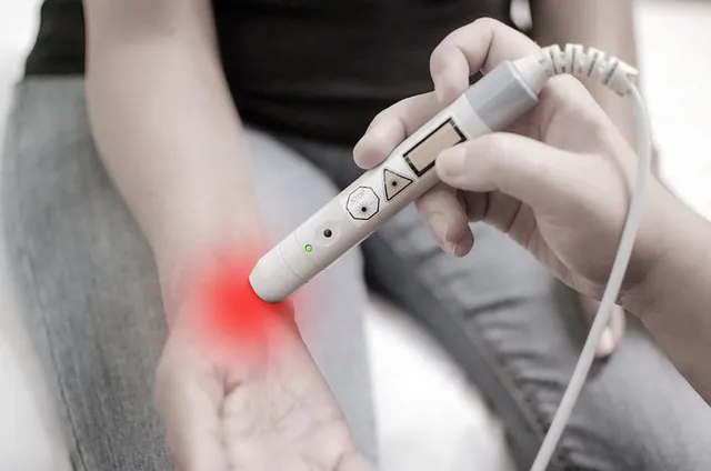
Why Chronic Inflammation Is a Silent Process
- Different cellular actors: acute inflammation is primarily driven by neutrophils — fast-moving white blood cells that arrive early and resolve quickly. Chronic inflammation is primarily maintained by macrophages and T-lymphocytes — longer-lived cells that set up residence in inflamed tissues and sustain inflammatory signaling indefinitely.
- Lower amplitude, longer duration: chronic inflammation produces TNF-alpha, IL-6, and IL-1β at levels 2-5x above baseline — not the 10-100x spikes seen during acute infection or injury. Modest enough to sit below the threshold that triggers dramatic symptoms, but enough for cumulative tissue damage across years.
- Tissue specificity determined by individual vulnerability: where chronic inflammation damages most depends on genetic predisposition, prior injuries, and the specific distribution of visceral fat and immune cell concentrations in the body. In one person, joint degeneration. In another, vascular damage. In a third, neuroinflammation and mood disruption. Same underlying process, different presentations.
Acute inflammation announces itself loudly: the swollen ankle, the hot infected wound, the fever. It’s designed to get attention and motivate protective behavior. Chronic systemic inflammation doesn’t work that way. It runs at a low enough intensity that the immune system never fully mobilizes its emergency response, and at a high enough persistence that tissues accumulate damage over years instead of days.
The cellular biology of chronic low-grade inflammation differs from acute inflammation in important ways:
This is why the symptom checklist matters: chronic inflammation doesn’t present uniformly. Several of these symptoms can show up without anyone recognizing the common thread. The Silent Inflammation Scorecard below helps assess total inflammatory burden — not just any single symptom in isolation.
Sign 1: Persistent Joint Pain and Stiffness (Without Specific Injury)
- Distribution: diet-driven inflammation tends to hit multiple large joints bilaterally (both knees, both hips, both hands); autoimmune arthritis follows characteristic patterns (RA: small joints of hands and feet, symmetrically; psoriatic arthritis: asymmetric, often with spine involvement)
- Blood markers: autoimmune conditions typically produce positive rheumatoid factor (RF), anti-CCP antibodies, or specific HLA markers; diet-driven inflammation typically shows elevated CRP/ESR without those specific autoantibodies
- Response to anti-inflammatory dietary changes: diet-driven joint inflammation often responds significantly within 4-8 weeks of dietary intervention; established autoimmune joint disease responds partially but usually needs additional treatment
Joint pain is the most commonly recognized symptom of inflammation, and also the most misattributed. Morning stiffness — particularly stiffness lasting more than 30-45 minutes before joints “warm up” — is a classic sign of inflammatory joint disease. Different from the brief stiffness that clears within minutes of movement, which is more characteristic of osteoarthritis mechanics.
Chronic systemic inflammation reaches joints through synovial fluid — the lubricating fluid inside joint capsules. Inflammatory cytokines (TNF-alpha and IL-1β particularly) trigger synovial membrane inflammation, driving excess fluid production, cartilage degradation, and joint space narrowing over time. The same cytokines produced by visceral fat, gut dysbiosis, and dietary inflammatory inputs that cause systemic effects reach joint tissue through circulation too.
What separates diet-and-lifestyle-driven joint inflammation from autoimmune joint disease:
Joint pain accompanied by morning stiffness over an hour, warmth or redness over the joints, or swelling — get a rheumatological workup before assuming it’s dietary in origin.
Sign 2: Brain Fog and Cognitive Slowness
- Worse after inflammatory meals (high sugar, restaurant food, alcohol the night before)
- Better in the morning, worse in the afternoon (correlating with cumulative dietary intake through the day)
- Improves significantly during periods of clean eating (fasting, travel to places with different food, anti-inflammatory dietary periods)
- Accompanied by other systemic inflammatory symptoms (joint pain, fatigue, gut issues)
Brain fog is maybe the most underappreciated inflammatory symptom — partly because it’s subjective and hard to measure, partly because modern culture has normalized cognitive impairment as a byproduct of busyness, stress, or age. “Can’t think straight today” gets accepted as a lifestyle complaint rather than investigated as a symptom.
The neurobiological mechanism is established: inflammatory cytokines produced peripherally (gut, visceral fat, joints) cross the blood-brain barrier — or signal across it through the vagus nerve and circumventricular organs — and activate microglia, the brain’s resident immune cells. Activated microglia produce their own inflammatory cytokines locally, alter neurotransmitter metabolism, and reduce synaptic plasticity. This is neuroinflammation, and it produces measurable cognitive deficits across multiple experimental models.
Specifically, inflammatory cytokines — IL-1β and TNF-alpha especially — alter serotonin and dopamine metabolism by upregulating indoleamine 2,3-dioxygenase (IDO), an enzyme that diverts tryptophan away from serotonin synthesis toward quinolinic acid, a neurotoxic compound. Which is one reason systemic inflammation shows up linked to both depression and cognitive impairment — the same cytokine elevation depletes precursors to mood-regulating neurotransmitters while producing neurotoxic byproducts on the side.

Sign 3: Unexplained Fatigue
Fatigue has many causes — thyroid dysfunction, anemia, sleep apnea, depression, overtraining. Inflammatory fatigue has a distinctive quality experienced practitioners recognize: sleep doesn’t relieve it (unlike simple sleep deprivation), it worsens after inflammatory meals, and it coexists with other inflammatory symptoms rather than showing up alone.
The mechanism: inflammatory cytokines activate the “sickness behavior” pathway — an evolutionarily conserved response to infection producing fatigue, social withdrawal, reduced motivation, and increased sleep. Adaptive during acute infection, since it conserves energy for immune function. In chronic low-grade inflammation, the same pathway stays chronically partly activated, producing persistent but non-dramatic fatigue, reduced motivation, and decreased capacity for effortful activity.
IL-6 is particularly implicated in inflammatory fatigue. Elevated IL-6 shows up in patients with cancer-related fatigue, post-viral fatigue syndromes, and multiple sclerosis fatigue — all conditions where inflammatory mechanisms are well-established drivers of the fatigue symptom. In metabolic syndrome and obesity — both high-IL-6 states — fatigue is nearly universal.
The clinical test: does the fatigue respond to anti-inflammatory dietary intervention? Many people report that within 2-3 weeks of cutting seed oils and added sugar, afternoon energy normalizes noticeably — before any body composition changes, before any dramatic weight loss. This functional response is one of the more reliable signals that inflammatory mechanisms are contributing to fatigue.
Sign 4: Skin Manifestations
Skin is one of the more reliable readouts of systemic inflammatory status — not because it’s the most affected tissue, but because it’s visible and its changes are easy to track directly.
Several skin conditions have well-established inflammatory mechanisms:
Acne: P. acnes bacteria trigger TLR2 and TLR4 receptors on sebocytes and keratinocytes, activating NF-kB and driving IL-1β production. Dietary factors that amplify this pathway — high glycemic index carbohydrates (driving IGF-1 and mTOR → sebum production) and dairy (skim milk particularly, through IGF-1 pathways) — have growing clinical support behind them. Multiple RCTs have found low-glycemic dietary interventions significantly reduce acne lesion counts.
Eczema/atopic dermatitis: a Th2-dominant inflammatory condition with IL-4, IL-13, and IL-31 as primary mediators. Systemic dietary inflammation can worsen atopic conditions by amplifying the Th2 response. Gut microbiome dysbiosis is increasingly recognized as a driver of atopic dermatitis — early-life antibiotic use, low-fiber diet, and low microbiome diversity are associated with higher eczema risk.
Psoriasis: a Th17-driven autoimmune condition producing excess keratinocyte proliferation. Systemic inflammatory load from obesity, metabolic syndrome, and poor diet clearly worsens psoriasis severity — dermatology classifies the condition as an “inflammatory disease,” not just a skin condition. Anti-inflammatory dietary interventions produce meaningful improvement in psoriasis severity scores across multiple trials.
General skin dullness, early aging, and poor wound healing: less specific, but real. Chronic inflammation accelerates skin AGE formation (reducing elasticity), reduces collagen synthesis (through MMP upregulation), and impairs the wound-healing cascade. The “skin aging diet” — high sugar, refined carbs, seed oils — is mechanistically the pro-inflammatory diet, via AGE formation and lipid oxidation.
Sign 5: Weight Gain and Difficulty Losing Weight

Inflammation drives weight gain through:
- Insulin resistance: TNF-alpha directly impairs insulin signaling by serine-phosphorylating IRS-1 (insulin receptor substrate 1), reducing glucose uptake in muscle and driving preferential fat storage in adipose tissue
- Leptin resistance: inflammatory cytokines impair leptin signaling in the hypothalamus — the brain’s satiety signaling system — making fullness harder to detect and increasing food intake
- Altered gut microbiome: dysbiosis shifts the microbiome toward species that extract more calories from food and produce LPS that drives hepatic fat accumulation
- Sleep disruption: inflammatory cytokines impair sleep quality; poor sleep increases ghrelin (hunger hormone) and decreases peptide YY (satiety hormone), increasing caloric intake by 200-400 calories a day on average in sleep-deprived individuals
Weight gain drives inflammation through:
- Visceral adipose tissue secreting TNF-alpha, IL-6, and resistin continuously
- Hypoxia in enlarged fat cells triggering HIF-1α (hypoxia-inducible factor) and inflammatory signaling
- Adipocyte death in rapidly expanding fat tissue releasing cell-free DNA and DAMPs (damage-associated molecular patterns) that activate inflammasomes
The practical implication: the inflammation doesn’t fully resolve without addressing the weight, and the weight doesn’t easily resolve without addressing the inflammation. Dietary quality, not caloric restriction, is the better entry point — reducing inflammatory inputs breaks the cycle from the inflammation side, which then opens the door to metabolic improvement and weight loss.
Sign 6: Digestive Problems
- Bloating that’s worse after sugar, refined carbs, or restaurant meals (consistent with dysbiosis fed by inflammatory foods)
- Alternating gut function that correlates with dietary patterns over the preceding 48 hours
- Gut symptoms that improve dramatically during travel (often involving food changes rather than just location changes)
- Resolution with anti-inflammatory dietary intervention in 2-4 weeks
Bloating, gas, alternating constipation and loose stools, discomfort after eating, a general sense of digestive unpredictability — extremely common inflammatory symptoms routinely written off as irritable bowel syndrome (IBS) or “sensitive stomach” without any real investigation of inflammatory mechanisms behind them.
The gut is both a major site of inflammatory pathology and a generator of systemic inflammatory signals. Intestinal permeability (“leaky gut”) lets bacterial products enter circulation; gut dysbiosis reduces anti-inflammatory SCFA production; intestinal macrophage activation from dietary inflammatory inputs produces local and systemic inflammatory signaling. Digestive symptoms can be either the cause or the consequence of chronic systemic inflammation — usually both, at the same time.
Low-grade digestive inflammation patterns with an inflammatory basis:
Signs 7-11: Additional Markers of Systemic Inflammation
Sign 7: Frequent illness (more than 3-4 viral infections per year in adults). Chronic inflammation produces immune dysfunction that impairs antiviral response. Counter-intuitively, a chronically activated immune system in an inflammatory state gets less effective at targeted viral response — resources are tied up managing the persistent inflammatory signaling, leaving less capacity for acute immune responses. Elevated TNF-alpha and IL-6 can actually impair natural killer cell function and interferon signaling — the front-line antiviral defenses.
Sign 8: Sleep disruption and non-restorative sleep. NF-kB and inflammatory cytokines regulate circadian rhythm and sleep architecture. Elevated TNF-alpha shifts sleep toward lighter stages and increases nighttime awakening. The correlation runs both directions: poor sleep elevates inflammatory cytokines, and elevated cytokines impair sleep quality. Breaking the cycle — dietary anti-inflammatory intervention plus sleep hygiene — is one of the faster ways to demonstrate inflammatory status improvement subjectively.
Sign 9: Depression and mood instability. The inflammatory hypothesis of depression isn’t fringe theory anymore — it’s increasingly mainstream psychiatric research. Roughly one-third of people with major depressive disorder have elevated CRP and other inflammatory markers. The tryptophan metabolism pathway described earlier (the IDO enzyme diverting tryptophan toward quinolinic acid under inflammatory conditions) provides a direct biochemical link between systemic inflammation and depressive symptoms. Which is why anti-inflammatory dietary interventions show efficacy in depression trials — they address a genuine biological mechanism, not just a lifestyle variable.
Sign 10: Slow recovery from exercise or injury. Inflammatory resolution requires specific pro-resolving mediators (resolvins, protectins, maresins) derived from omega-3 fatty acids. Omega-3 insufficiency combined with excess omega-6 impairs production of these resolution signals, prolonging inflammatory states after both exercise-induced muscle damage and physical injury. Muscles staying sore 4-5 days after training (versus 1-2 days in well-nourished athletes), or injuries healing unusually slowly — omega-3 insufficiency and chronic inflammatory burden are likely contributors.

The Silent Inflammation Scorecard
Use this scorecard to assess inflammatory burden. Score 1 point for each symptom present to a significant degree — not occasionally, but persistently over the past 3+ months:
- Joint pain or stiffness (morning stiffness lasting more than 30 minutes) — 1 point
- Brain fog, mental slowness, or poor concentration most afternoons — 1 point
- Fatigue not relieved by adequate sleep — 1 point
- Skin issues (acne, eczema, psoriasis, or persistent dullness/rash) — 1 point
- Weight gain in the past 2 years without major dietary changes, or significant difficulty losing weight despite effort — 1 point
- Digestive discomfort, bloating, or irregular bowel function most weeks — 1 point
- 4 or more viral infections per year — 1 point
- Non-restorative sleep or frequent nighttime awakening — 1 point
- Persistent low mood, anxiety, or mood instability — 1 point
- Slow exercise recovery (muscle soreness lasting 4+ days) or slow wound healing — 1 point
- Gum disease, frequent cavities, or oral health issues — 1 point
Score 0-2: Low inflammatory burden. Focus on maintenance through foundational anti-inflammatory dietary habits.
Score 3-5: Moderate inflammatory burden. Significant dietary intervention warranted. Track hs-CRP. Implement the Inflammation Elimination Protocol fully.
Score 6-8: High inflammatory burden. Consider comprehensive biomarker testing (hs-CRP, fasting insulin, ferritin, omega-3 index). Dietary intervention should be the top health priority. Consider consultation with a functional medicine practitioner who can work with you on root causes.
Score 9-11: Very high inflammatory burden. Rule out autoimmune conditions and other medical causes. Comprehensive lifestyle and dietary intervention warranted. Physician partnership important.
“The body keeps score. It doesn’t send you a memo — it changes function. By the time you’re paying attention, the problem has usually been building for years. The symptoms are the message; the inflammation is the language.” — Adapted from systemic inflammation research literature
Lab Tests Worth Requesting
Subjective symptoms are an imprecise tool. These lab tests provide objective quantification of inflammatory status and can track improvement over time:
hs-CRP (high-sensitivity C-reactive protein): the most clinically validated marker of systemic inflammation. Produced by the liver in response to IL-6. Optimal: below 1.0 mg/L. Low risk: 1.0-3.0 mg/L. High risk: above 3.0 mg/L. Critical note: hs-CRP also rises with acute infections. Test after 4 weeks clear of illness for an accurate chronic inflammation reading.
ESR (erythrocyte sedimentation rate): measures how quickly red blood cells settle in a test tube — faster settling means more fibrinogen and other inflammatory proteins in the blood. Less specific than hs-CRP but a useful secondary marker, particularly for monitoring autoimmune conditions. Normal: under 15 mm/hr for men under 50, under 20 mm/hr for women under 50.
Ferritin: an iron-storage protein and acute-phase reactant. Elevated ferritin (above 200 ng/mL in women, above 300 ng/mL in men) without iron overload or a recent blood transfusion is a reliable indicator of chronic inflammation. Also useful for ruling out iron deficiency anemia as a cause of fatigue.
Fasting insulin: not a direct inflammatory marker, but insulin resistance is both a cause and consequence of systemic inflammation, making it a valuable metabolic context marker. Optimal: below 5 μIU/mL. Moderate insulin resistance: 10-20 μIU/mL. Significant insulin resistance: above 20 μIU/mL.

HbA1c: three-month average blood glucose. Optimal: below 5.3%. Pre-diabetic range begins at 5.7%. Quantifies the degree of chronic glucose dysregulation, which independently drives AGE formation and NF-kB activation.
Omega-3 Index: the percentage of EPA + DHA in red blood cell membranes, reflecting the past 3-month dietary omega-3 status. Optimal: 8% or above. Below 4% signals significant deficiency. Not routinely ordered by most physicians, but available through specialty labs and home testing kits (OmegaQuant). Knowing the baseline omega-3 index helps calibrate how aggressively to supplement or emphasize dietary fatty fish.
When Symptoms Warrant Immediate Medical Evaluation
Lifestyle-driven chronic inflammation is a real and important health issue that dietary intervention meaningfully addresses. That said, some symptom combinations warrant medical evaluation to rule out specific inflammatory diseases requiring different treatment:
- Joint pain + significant morning stiffness lasting over an hour + symmetrical small joint involvement: rule out rheumatoid arthritis (rheumatologist referral)
- Fatigue + significant unexplained weight gain or loss + cold intolerance or heat intolerance: rule out thyroid disease (TSH, free T3, free T4)
- Gut symptoms + blood in stool or significant unintentional weight loss: rule out inflammatory bowel disease and colorectal pathology
- Skin rash + joint pain + sun sensitivity: rule out lupus (ANA panel)
- Persistent elevated CRP (above 5-10 mg/L) without dietary explanation: evaluate for occult infection, malignancy, or autoimmune disease
- Fatigue + night sweats + swollen lymph nodes: needs medical evaluation to rule out lymphoma and other malignancies
The dietary anti-inflammatory protocol is a powerful intervention for lifestyle-driven systemic inflammation. It is not a substitute for medical diagnosis of specific conditions that may be driving symptoms.
Chronic Inflammation Symptoms Q&A
Q: Can I have high inflammation with a normal CRP?
Yes. CRP reflects systemic inflammatory burden but may stay normal in localized inflammation (a single inflamed joint, say, with limited systemic spillover) or in early inflammatory conditions that haven’t yet crossed the threshold triggering hepatic CRP production. CRP is also a relatively insensitive marker for neuroinflammation — brain inflammation can be significant without proportional systemic CRP elevation. Other markers (ferritin, fasting insulin, homocysteine) fill in complementary information. Ultimately, multiple symptoms from the scorecard above raise the likelihood of inflammation regardless of CRP — inflammatory biomarkers are supporting evidence, not definitive proof.
Q: How quickly can CRP drop with dietary changes?
Clinical evidence shows 20-40% reductions in CRP achievable within 4-8 weeks of consistent anti-inflammatory dietary intervention. The magnitude of improvement correlates with the magnitude of dietary change and baseline inflammatory status. People with the highest baseline CRP typically show the most dramatic reductions. CRP reductions of 50% or more over 12-16 weeks show up in studies using comprehensive lifestyle interventions (diet + exercise + sleep improvement together).
Q: My doctor says my inflammation markers are “normal.” Should I still pursue this protocol?
Standard lab “normal ranges” are built on population averages, which include the highly inflamed general population as part of the reference group. A CRP of 2.8 mg/L may be “within range” but still carries significantly higher cardiovascular risk than a CRP below 1.0 mg/L. The optimal targets listed above trace to cardiovascular and longevity outcome data, not population averages. “In range” and “optimal” simply aren’t the same thing.

The evidence increasingly suggests a meaningful inflammatory component in a subset of CFS/ME cases — particularly those with post-infectious onset, where viral-triggered immune dysfunction may have established a self-sustaining inflammatory state. Elevated inflammatory markers have shown up across multiple CFS/ME cohorts. CFS/ME is a heterogeneous condition with multiple possible underlying mechanisms, though, and anti-inflammatory dietary intervention is not a cure — it may be one supportive element within a broader management approach.
Q: Can inflammation cause weight loss rather than weight gain?
Yes, in acute and severe inflammatory states. High-grade inflammation from cancer, severe infection, or advanced autoimmune disease typically causes weight loss (cachexia) through elevated TNF-alpha (historically called “cachectin” for this reason) and IL-6, which suppress appetite and increase metabolic rate. The weight gain association is characteristic of the lower-grade chronic metabolic inflammation driven by dietary factors and obesity — not the high-grade inflammation of serious disease. Unintentional weight loss should always prompt medical evaluation.
Q: Are there genetic variants that make some people more susceptible to dietary inflammation?
Yes. Several genetic polymorphisms affect inflammatory response to dietary inputs. TNF-alpha promoter polymorphisms affect baseline TNF production. IL-6 gene variants affect inflammatory cytokine levels. FADS1/FADS2 polymorphisms affect omega-6 to omega-3 conversion efficiency. APO E4 genotype affects both cardiovascular and neurological inflammatory risk from dietary fat. None of this determines destiny — dietary quality powerfully modifies expression of inflammatory gene variants — but it does explain why some people run more sensitive than others to the same dietary insults.
The Autoimmune Boundary: When Inflammation Becomes Self-Attack
- Lifestyle-driven chronic inflammation may lower the threshold for autoimmune triggering in genetically susceptible individuals. Chronic NF-kB activation from poor diet and gut dysbiosis creates an immune environment where self-tolerance mechanisms are more easily overwhelmed.
- Once established, autoimmune conditions get significantly worsened by lifestyle inflammatory inputs. The same dietary factors driving systemic inflammation in healthy people amplify autoimmune disease activity in people with established conditions.
- The gut microbiome is increasingly recognized as a key modifier of autoimmune risk and disease activity. Specific gut microbiome patterns are associated with rheumatoid arthritis, multiple sclerosis, and lupus. Dietary interventions shifting microbiome composition away from dysbiotic patterns may modulate autoimmune disease activity.
Chronic systemic inflammation from lifestyle factors and autoimmune inflammation sit on a spectrum, not as categorically separate conditions. Understanding the relationship clarifies both the limits of lifestyle intervention and the opportunities within those limits.
Autoimmune conditions involve the immune system misidentifying the body’s own tissues as foreign and mounting inflammatory attacks against them. The classical autoimmune diseases — rheumatoid arthritis (attacks joint synovium), Hashimoto’s thyroiditis (attacks thyroid tissue), lupus (attacks multiple tissues), Type 1 diabetes (attacks pancreatic beta cells), psoriasis (attacks skin), multiple sclerosis (attacks myelin in the central nervous system) — have clear autoimmune mechanisms with identifiable autoantibodies or autoreactive T cells.
The relationship between lifestyle-driven inflammation and autoimmune disease runs bidirectional:
For anyone with an established autoimmune condition, the lifestyle anti-inflammatory protocol stays highly relevant but functions as a disease-modifying adjunct, not a cure. Multiple clinical trials have shown meaningful improvements in disease activity scores for rheumatoid arthritis, psoriasis, and inflammatory bowel disease from anti-inflammatory dietary interventions — improvements that reduce symptom burden and may reduce medication requirements, without eliminating the need for ongoing medical management of the underlying autoimmune condition.
The specific additional dietary considerations for autoimmune conditions:
- The Autoimmune Protocol (AIP): a more restrictive elimination protocol removing all grains, legumes, nightshades, eggs, dairy, nuts, seeds, alcohol, and NSAIDs for 30-90 days, then systematically reintroducing foods to identify individual immune triggers. AIP has pilot study data showing reduced disease activity in Hashimoto’s thyroiditis and inflammatory bowel disease. Not appropriate as a first-line general population approach — the restriction is significant and the evidence base is limited — but for treatment-resistant autoimmune conditions, it provides a structured framework for identifying individual dietary triggers.
- Elimination and reintroduction for specific triggers: even without full AIP, systematic elimination of the most commonly reactive foods in autoimmune conditions — dairy (A1 casein), gluten (in celiac and non-celiac gluten sensitivity), nightshade alkaloids (for arthritis), and eggs (for some eczema presentations) — followed by structured reintroduction, can identify individual food triggers worth permanently avoiding.
- Gut permeability repair: in autoimmune conditions with clear gut dysbiosis or intestinal permeability (IBD, autoimmune thyroid disease), targeted gut healing interventions including bone broth, collagen peptides, zinc carnosine, and L-glutamine may provide additional benefit in restoring gut barrier function beyond what dietary quality improvement alone achieves.
Testing Your Inflammatory Status Over Time: A Monitoring Framework
One of the more empowering aspects of addressing chronic inflammation is that the biomarkers quantifying it are accessible, inexpensive, and change measurably with intervention. A monitoring framework turns an anti-inflammatory lifestyle from an act of faith into a data-driven practice.
The core monitoring protocol:
At baseline (before starting any dietary intervention): hs-CRP, fasting insulin, ferritin, HbA1c, and waist circumference. Optionally: Omega-3 Index, to quantify current omega-3 status and track response to dietary fish and supplementation changes. These five markers provide a comprehensive snapshot of current inflammatory and metabolic status. Record everything with dates.
At 3 months: retest hs-CRP, fasting insulin, and HbA1c. These are the fastest-moving markers — three months of consistent dietary change should produce measurable change if the intervention is working. hs-CRP improving but insulin and HbA1c not? Residual carbohydrate issues may need addressing. Insulin improving but hs-CRP still elevated? Focus on non-dietary inflammatory inputs — sleep, stress, dental health, possible occult infection.
At 6 months: full panel repeat including ferritin and Omega-3 Index. By this point, visceral fat changes are often measurable, reflected in ferritin normalization and waist circumference reduction. The Omega-3 Index gives a 3-6 month picture of fatty acid status changes.
Ongoing (annually or semi-annually): hs-CRP as the primary maintenance marker. Full panel annually to track long-term trajectory. Most people maintaining the dietary protocol maintain or continue improving their biomarker profile over 12-24 months.
The psychological value of biomarker tracking extends beyond the clinical information it provides: it creates an objective feedback loop that makes dietary adherence feel purposeful rather than arbitrary. Watching hs-CRP drop from 2.8 to 1.1 over 12 weeks makes the dietary discipline required to get there feel proportionate to a concrete outcome. This closes the behavioral loop in a way subjective symptom tracking — susceptible to placebo effects and daily variability — can’t.
The Compound Effect: Why Treating Each Symptom Separately Fails
The most important insight in this entire guide is the one embedded in Karen’s story at the start: treat each symptom of chronic inflammation as an isolated problem, and the result is a collection of band-aids over a single wound that never heals. The joint pain gets NSAIDs that mask the symptom while the underlying inflammatory load keeps building. The fatigue gets caffeine that drives the adrenal system harder while the root cause goes unaddressed. The brain fog gets attributed to stress and managed with anxiety medications doing nothing for the neuroinflammation underneath it. The skin issues get topical steroid creams that suppress local inflammation without touching the systemic driver.
Every year this approach continues, the inflammatory burden deepens, tissue damage accumulates, and more symptoms show up to be treated in isolation. Not a criticism of the individual physicians who prescribed these treatments — they were working within a symptom-management model that doesn’t have good tools for identifying and addressing systemic root causes inside a standard clinic visit. It’s an observation that the model itself is inadequate for the problem of chronic systemic inflammation.
The alternative model is what this guide outlines: assess the total inflammatory burden using the scorecard and biomarkers, identify root causes systematically (diet, sleep, gut health, stress, visceral fat), and address them in priority order using the protocol framework. Do this, and something remarkable often happens — multiple seemingly unrelated symptoms improve simultaneously, because they were never truly separate problems in the first place.
Karen’s joints improved. Her brain fog lifted. Her energy came back. Her skin cleared. She lost twelve pounds without trying particularly hard. Not twelve separate medical victories. One inflammation victory, showing up in twelve different tissue systems. That’s the compound effect of root cause thinking applied to chronic inflammation. Accessible to anyone willing to do the dietary work and track the results honestly.
Once you’ve identified your inflammatory burden with this scorecard, the full dietary protocol for addressing it is in the Anti-Inflammatory Diet Plan. For understanding what your CRP number actually means and what to do with the result, see the CRP Test guide.
References: Ridker PM. Clinical application of C-reactive protein for cardiovascular disease detection and prevention. Circulation. 2003. | Slavich GM, Irwin MR. From stress to inflammation and major depressive disorder. Psychol Bull. 2014. | Esposito K et al. Effect of Mediterranean-style diet on endothelial dysfunction. JAMA. 2004. | Calder PC et al. Dietary factors and low-grade inflammation in relation to overweight and obesity. Br J Nutr. 2011.
The Practical Framework: Applying Chronic Inflammation Symptoms Signs In Real Life
Editorial StandardsCorrectionsMedical DisclaimerAbout Our ContentAffiliate DisclosureSite Map