
The Scan That Nobody Expected
Carlos had been tired for two years. Not the tired from poor sleep or a hard week — the steady, pervasive fatigue that makes everything slightly harder than it should be. His doctor had run bloodwork twice. Both times, his liver enzymes came back “mildly elevated” — ALT at 58, AST at 45, GGT at 72. “Worth watching,” his doctor said. “Come back in six months.” No explanation for what might be causing it. No dietary guidance. No discussion of what those numbers meant or what could be done about them.
Two years of “worth watching” later, a different physician ordered an ultrasound. It showed moderate non-alcoholic fatty liver disease (NAFLD) — liver fat deposition in approximately 20-30% of his liver. Carlos had NAFLD and nobody had told him. Not because the physicians were negligent, but because “elevated liver enzymes” in the absence of alarming numbers often sits in a clinical holding pattern — monitored, not investigated. He wasn’t drinking excessively. He wasn’t taking hepatotoxic medications. His enzymes were elevated, just not dramatically so. The system didn’t trigger on him, and his liver quietly accumulated fat for two years while he was told to come back in six months.
The liver is the most metabolically complex organ in the body — a chemical factory processing everything you eat, drink, breathe, and metabolize. When it’s struggling, the signs are often subtle, the standard tests are often inadequate, and the clinical system often waits until the problem is significant before responding with anything beyond monitoring. This article covers what to look for, what to test, and what to do — specifically for the large and growing population whose livers are accumulating damage from metabolic and inflammatory drivers that standard medical panels are poorly designed to catch early.
What the Liver Actually Does: Why It Matters So Much
Glucose metabolism: the liver is the primary organ regulating blood glucose homeostasis. After a meal, it captures glucose from the portal circulation, converts it to glycogen for storage (glycogenesis), or converts excess to fat (de novo lipogenesis — the primary mechanism of fatty liver development in fructose-overconsumption). During fasting, the liver releases glucose from glycogen stores (glycogenolysis) and produces new glucose from non-carbohydrate precursors (gluconeogenesis). When the liver is fatty and inflamed, these processes get dysregulated — glycogen storage impairs, gluconeogenesis runs overactive, de novo lipogenesis runs excessive. This creates the insulin resistance pattern that drives metabolic syndrome.
Lipid metabolism: the liver synthesizes cholesterol, triglycerides, and phospholipids; packages them into lipoproteins for transport; and processes dietary lipids from chylomicron remnants. It’s also the primary site of beta-oxidation (fat burning) when energy demands require it. A fatty liver is often a liver where beta-oxidation has become impaired — it stores fat rather than burning it, partly because the lipid accumulation itself impairs the mitochondrial function efficient fat oxidation requires.
Detoxification: the liver’s cytochrome P450 enzyme system metabolizes virtually every drug, environmental toxin, and endogenous waste product that needs converting to a water-soluble form for excretion. The two-phase detoxification process — Phase I (oxidation via cytochrome P450) and Phase II (conjugation to increase water solubility) — requires specific nutrients at each phase. When liver function is impaired, detoxification capacity drops, contributing to the systemic toxin accumulation behind the fatigue and cognitive fog common in liver disease.
Protein synthesis: the liver produces most of the proteins circulating in the blood, including albumin (osmotic regulator), clotting factors (prothrombin, fibrinogen), acute-phase reactants (CRP, ferritin, alpha-1-antitrypsin), and transport proteins. This protein synthesis function is one of the last to be impaired in progressive liver disease — meaning by the time albumin and clotting factors measure low, significant liver damage has already occurred.
Bile production: the liver produces bile — a mixture of bile salts, cholesterol, bilirubin, and phospholipids — essential for fat-soluble vitamin absorption (A, D, E, K), fat digestion, and eliminating cholesterol and waste metabolites from the body. Bile also plays a significant role in gut microbiome regulation — bile acids are antimicrobial in the small intestine and modulate microbial community composition throughout the gut. Impaired bile production and flow (cholestasis) creates secondary problems in fat digestion, vitamin absorption, and gut microbiome health.
Reading Liver Blood Tests: ALT, AST, GGT, and Beyond
Standard liver panels measure aminotransferases and other hepatic enzymes. Understanding what each marker reflects, and what its elevation actually means, moves someone from passive recipient of “slightly elevated, worth watching” to active investigator of their liver’s functional status.
ALT (Alanine Aminotransferase) is the most liver-specific of the standard enzymes. Found primarily in liver cells, and its elevation in the bloodstream indicates hepatocyte damage — liver cell death or disruption releasing intracellular enzymes into the blood. The standard normal range is approximately 7-56 U/L for men and 7-35 U/L for women (lab ranges vary). But research by Prati and colleagues established that the upper limit of normal for ALT in a metabolically healthy population runs significantly lower — approximately 30 U/L for men and 19 U/L for women. “Normal” ALT up to 56 U/L includes people with significant early NAFLD, and the higher the “normal” threshold sits, the more early liver disease slips through undetected.
AST (Aspartate Aminotransferase) is less liver-specific than ALT — it’s also found in heart muscle, skeletal muscle, kidneys, and red blood cells. Isolated AST elevation without ALT elevation typically points to a non-liver source (muscle injury from intense exercise is a common benign cause). When both ALT and AST are elevated, liver involvement is more likely. The AST:ALT ratio adds diagnostic information: a ratio above 2:1 is associated with alcoholic liver disease; a ratio below 1:1 (ALT higher than AST) is more common in NAFLD.
GGT (Gamma-Glutamyl Transferase) is the liver enzyme with the strongest association with alcohol consumption and with metabolic disease. GGT is induced by alcohol (a reliable marker of even moderate alcohol use), by insulin resistance, by fatty liver, and by certain medications. It’s also sensitive to oxidative stress in the liver — elevated GGT reflects the liver’s depleted glutathione reserves in response to oxidative challenge. GGT elevation alongside normal or mildly elevated ALT and AST, in someone who doesn’t drink heavily, is a strong signal of metabolic liver stress. Optimal GGT sits below 25 U/L for men and below 18 U/L for women — far below the typical lab “normal” upper limit of 55-80 U/L.
Alkaline Phosphatase (ALP) is elevated in cholestatic conditions — when bile flow is obstructed or impaired. Also elevated in bone disease, which is why elevated ALP without elevated GGT often points to bone rather than liver. Isolated ALP elevation alongside elevated GGT indicates biliary dysfunction — impaired bile production or flow — rather than hepatocellular damage.
Bilirubin is the breakdown product of heme from red blood cell destruction, processed by the liver and excreted in bile. Elevated total bilirubin with elevated direct (conjugated) bilirubin indicates impaired bile excretion. Mildly elevated indirect (unconjugated) bilirubin on its own is common in Gilbert’s syndrome — a benign genetic condition affecting bilirubin conjugation that affects approximately 5% of the population and needs no treatment.
Albumin and Prothrombin Time (PT/INR) reflect the liver’s protein synthesis function. These markers stay normal in early and moderate liver disease; their impairment signals advanced hepatic dysfunction with reduced synthetic capacity. Normal albumin and PT/INR do not rule out significant early liver disease — they’re late-stage markers, not early warning signals.
Non-Alcoholic Fatty Liver Disease: The Epidemic Nobody Talks About
NAFLD is the most common liver condition worldwide, affecting an estimated 25% of the global adult population — approximately 2 billion people. In the United States, prevalence estimates range from 30-40% of adults, with rates rising as obesity, metabolic syndrome, and high-fructose dietary patterns keep expanding. A health crisis of enormous scale, receiving a tiny fraction of the public health attention devoted to conditions affecting far fewer people.
NAFLD exists on a spectrum: simple steatosis (fat accumulation without significant inflammation) at one end, non-alcoholic steatohepatitis (NASH — fat plus inflammation and fibrosis) in the middle, cirrhosis at the severe end. Progression from simple steatosis to NASH and cirrhosis isn’t inevitable — perhaps 20-30% of NAFLD patients develop NASH — but the factors driving progression are precisely the factors most characteristic of the modern metabolic environment: insulin resistance, fructose overconsumption, sedentary lifestyle, gut dysbiosis.
Fructose metabolism is particularly important to understand here. Fructose — from table sugar (sucrose), high-fructose corn syrup, fruit juice, and certain fruits consumed in excess — is metabolized almost exclusively by the liver (unlike glucose, which any cell can use). When fructose arrives at the liver in amounts exceeding its capacity to oxidize it or convert it to glycogen, de novo lipogenesis converts it directly to fat. This is the primary dietary pathway to NAFLD: the liver essentially converts fructose intake into liver fat in real time.
The standard clinical message that NAFLD is primarily a weight problem is incomplete. Thin people develop NAFLD too (the “TOFI” pattern — thin outside, fat inside — where metabolically unhealthy people at normal weight carry excess visceral and liver fat). High fructose intake drives NAFLD even in lean people with normal BMI. Sedentary lifestyle impairs the fat oxidation that would otherwise clear liver fat. Gut dysbiosis — particularly LPS translocation from a compromised gut barrier — directly activates Kupffer cells in the liver, driving the hepatic inflammation that converts simple steatosis to NASH. The full NAFLD picture requires understanding diet, body composition, exercise, and gut health all at once.
Milk Thistle, NAC, and the Evidence for Liver Support
Several supplements have meaningful clinical evidence for supporting liver health, worth knowing about particularly for people with elevated liver enzymes or confirmed NAFLD who are implementing lifestyle changes and want additional support for the repair process.
Milk thistle (silymarin) is the most widely studied hepatoprotective supplement. Silymarin — the active complex of flavonolignans extracted from milk thistle seeds — has antioxidant, anti-inflammatory, and antifibrotic properties in the liver. It reduces lipid peroxidation in hepatocytes, modulates inflammatory cytokine production in Kupffer cells, and has been shown to directly inhibit collagen synthesis in hepatic stellate cells (the cells responsible for liver fibrosis). Multiple randomized trials in NASH patients have documented improvements in ALT, AST, and liver histology with silymarin supplementation. A 2017 meta-analysis in the European Journal of Gastroenterology found significant ALT and AST reductions with silymarin across multiple NAFLD trials. Those trials used standardised extract in divided doses through the day rather than a single hit, and the phospholipid-bound form (silymarin-phosphatidylcholine complex) has markedly better bioavailability than plain extract — a detail that explains a good deal of the inconsistency between studies.
N-Acetylcysteine (NAC) is the direct precursor to glutathione — the primary antioxidant protecting hepatocytes from oxidative damage. The liver is the primary glutathione-producing organ in the body, and oxidative stress in NAFLD rapidly depletes hepatic glutathione reserves. NAC replenishes them, protecting against the oxidative hepatocyte damage that drives the progression from simple steatosis to NASH. NAC is used clinically in hospitals as the antidote for acetaminophen overdose (which causes liver failure specifically through glutathione depletion) — its hepatoprotective mechanism is well-established. The NAFLD literature on NAC sits well below the hospital acetaminophen protocols, which is worth knowing when reading about it. As a bonus, NAC also supports gut barrier integrity and has immune-modulating properties that address several of the gut-liver axis factors driving NAFLD.
Berberine has emerged as one of the most evidence-backed supplements for metabolic liver disease. It activates AMPK (AMP-activated protein kinase), the cellular energy sensor that promotes fat oxidation and inhibits fat synthesis — the exact pathways dysregulated in NAFLD. Multiple randomized trials comparing berberine to metformin for metabolic parameters in NAFLD patients have found comparable or superior effects on liver enzymes, liver fat content (measured by ultrasound), insulin resistance, and inflammatory markers. Those trials split the daily amount across meals rather than giving it all at once, largely because berberine’s gastrointestinal side effects cluster at single large doses. Berberine also modulates gut microbiome composition favorably — another mechanism relevant to NAFLD given the gut-liver axis connection.
The Liver Health Assessment Framework
The Liver Health Assessment is a tiered framework for evaluating liver status, identifying the likely drivers of any dysfunction, and implementing a targeted improvement protocol.
- Tier 1 — Standard Panel Optimization. Request ALT, AST, GGT, ALP, total and direct bilirubin, albumin. Evaluate against optimal (not just normal) ranges: ALT below 30 U/L (men), below 19 U/L (women); GGT below 25 U/L (men), below 18 U/L (women). Anything above these optimal thresholds warrants investigation of drivers rather than passive monitoring.
- Tier 2 — Advanced Liver Function. Add: fasting insulin and glucose (HOMA-IR calculation for insulin resistance); ferritin with iron studies (elevated ferritin with normal iron saturation indicates metabolic inflammation, a common NAFLD driver); triglycerides (above 150 mg/dL in fasting state suggests hepatic fat accumulation); and uric acid (elevated uric acid drives de novo lipogenesis through fructose metabolism by-product signaling).
- Tier 3 — Imaging If Indicated. Liver ultrasound is the standard first-line imaging for NAFLD assessment — it can detect liver fat accumulation with approximately 80% sensitivity when fat content exceeds 30%. Elastography (FibroScan) or liver MRI provide more precise fat content quantification and fibrosis staging if NASH is suspected. Indicated when ALT remains persistently elevated despite lifestyle intervention, or when clinical features (obesity, diabetes, elevated ferritin) suggest higher probability of advanced disease.
- Dietary Intervention. Primary: eliminate or dramatically reduce fructose — both from added sugars and fruit juice. Fructose is the primary dietary substrate for hepatic de novo lipogenesis and the most direct dietary driver of liver fat accumulation. Secondary: replace seed oils with olive oil and other stable fats — seed oil oxidation products contribute to hepatocyte oxidative stress. Tertiary: increase dietary fiber diversity (supports gut microbiome and reduces LPS translocation to the liver) and choline-rich foods (eggs, liver, seafood — choline is required for VLDL assembly that exports fat from the liver; choline deficiency directly causes fatty liver).
- Exercise Protocol. Both aerobic and resistance training reduce liver fat independent of weight loss. Aerobic exercise at 150+ minutes per week significantly reduces hepatic steatosis. Resistance training improves insulin sensitivity and reduces the hyperinsulinemia driving de novo lipogenesis. The combination beats either alone. Even absent weight loss, exercise consistently reduces liver fat content measurably within 8-12 weeks of regular training.
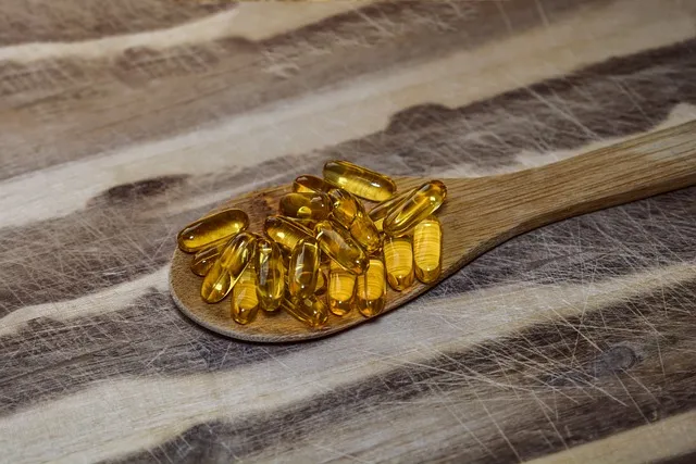
- Supplement Questions Worth Raising. Four compounds carry enough NAFLD evidence to be worth discussing with whoever is managing the liver panel: silymarin in the phospholipid-bound form, NAC, berberine where metabolic liver disease is confirmed, and choline where the diet is genuinely short of it. Omega-3 EPA/DHA belongs on that list too — multiple trials document liver fat reduction with omega-3 supplementation in NAFLD through inhibition of de novo lipogenesis and promotion of fat oxidation. What each of them is worth in a particular case depends on the panel, the diagnosis, and the medications already in play, which is a conversation rather than a shopping list.
“The liver is the most metabolically forgiving organ in the body — it can regenerate from surprisingly significant damage if the insults causing that damage are removed. But it cannot regenerate while the insults continue. The first intervention is always to stop doing the things that are causing the problem.”
The Gut-Liver Axis: Why Gut Health Drives Liver Health
One of the more important developments in liver disease understanding over the past decade is recognition of the gut-liver axis — the bidirectional communication pathway between gut microbiome and liver function — as a primary driver of NAFLD progression.
The portal vein drains all blood from the intestines directly to the liver. Everything passing from the gut into the bloodstream — nutrients, microbial metabolites, inflammatory molecules — reaches the liver first, before entering systemic circulation. That makes the liver the primary filter for gut-derived signals, and when the gut is producing abnormal signals, the liver is the first organ to feel the consequences.
In people with gut dysbiosis and elevated intestinal permeability, bacterial lipopolysaccharide (LPS) — the endotoxin from gram-negative bacterial cell walls — enters the portal circulation and reaches the liver in elevated concentrations. LPS binds to TLR4 receptors on Kupffer cells (liver macrophages), triggering a powerful inflammatory response: TNF-alpha, IL-6, IL-1β production in the liver that drives the hepatic inflammation characteristic of NASH. This LPS-driven Kupffer cell activation is one of the primary mechanisms distinguishing progressive NASH from simple steatosis — the inflammatory hit on top of the fat accumulation that drives fibrosis and disease progression.
This gut-liver axis connection explains why comprehensive NAFLD management has to include gut health interventions alongside the standard dietary and exercise recommendations. Reducing gut dysbiosis, supporting intestinal barrier integrity, and reducing LPS translocation directly protects the liver from the inflammatory signals driving progression. The post-antibiotic probiotic protocol, the gut-healing dietary pattern, and the bone broth elements described elsewhere in this series are as relevant to NAFLD management as they are to gut health specifically.
Common Questions About Liver Health Signs

Request a copy of the results with exact values. If ALT is above 30 U/L (men) or 19 U/L (women), or GGT is above 25 U/L (men) or 18 U/L (women), there’s a rational basis for pursuing the Tier 2 assessment described above — specifically fasting insulin, ferritin with iron studies, triglycerides, and uric acid. If those show metabolic liver disease features, request a liver ultrasound. This isn’t alarmism. It’s appropriate proactivity about a condition currently affecting roughly a third of American adults and significantly underdiagnosed at early stages.
Can NAFLD be reversed?
Yes, in the early stages (simple steatosis and mild NASH). Multiple studies have documented complete histological reversal of early NAFLD through lifestyle intervention — specifically the combination of dietary fructose reduction, regular aerobic exercise, and weight loss where applicable. The liver’s regenerative capacity is remarkable; it can clear fat accumulation and reverse early fibrosis if the metabolic insults driving the accumulation are removed. The more advanced the disease (significant fibrosis, cirrhosis), the less complete the reversal — which is why early identification and early intervention produces substantially better outcomes than waiting until enzymes are dramatically elevated.
Does alcohol cause the same kind of liver damage as NAFLD?
Alcoholic liver disease and NAFLD share many pathological features — fat accumulation, inflammation, fibrosis — through partially different mechanisms. Alcohol is directly hepatotoxic through its metabolism to acetaldehyde, which forms protein adducts that damage hepatocytes, deplete glutathione, and directly activate Kupffer cells. NAFLD is driven primarily by insulin resistance and fructose metabolism. In people with both NAFLD and significant alcohol use, the disease progresses much more aggressively — the two insults aren’t simply additive, they interact synergistically to accelerate hepatic damage. For people with elevated liver enzymes, even moderate alcohol consumption is a significant additional burden on an already-stressed liver.
What is choline, and how do I know if I’m getting enough?
Choline is an essential nutrient required for hepatic VLDL assembly — the process by which the liver packages fat for export into the bloodstream. Without adequate choline, the liver can’t efficiently export the fat it synthesizes or receives from the diet, and fat accumulates. Choline deficiency is actually a standard laboratory model for inducing NAFLD in animal studies — it reliably produces fatty liver when animals are fed choline-deficient diets. The best dietary choline sources are eggs (1 egg yolk provides approximately 150mg choline), liver (approximately 350mg per 3 oz), beef (approximately 115mg per 3 oz), and seafood (shrimp, 90mg per 3 oz). The adequate intake for adults is 425-550mg daily — achievable with eggs daily but insufficient on egg-free diets. Given that both NAFLD prevalence and low egg consumption have been rising simultaneously for decades, the choline gap in the population is significant and underappreciated.
Carlos’s resolution: After his NAFLD diagnosis, Carlos eliminated fructose from his diet (primarily sugar-sweetened beverages and fruit juices he hadn’t been counting), cut refined carbohydrates significantly, and began exercising five days a week — aerobic exercise three days, resistance training two. He added silymarin, NAC, and berberine at the protocol doses. At six months, his repeat liver ultrasound showed the fatty liver had completely resolved. His ALT was 22 U/L, his GGT was 18 U/L, and his fatigue — the original presenting symptom that started the whole investigation — had completely resolved. The liver had healed. It had needed only the right environment to do so.
Nutrients the Liver Needs to Function
Beyond the specific supplements discussed above, several nutrients are particularly critical for optimal liver function and are commonly deficient in populations with metabolic liver disease.
Choline, as discussed, is required for VLDL assembly and hepatic fat export. Methylation — the biochemical process of adding methyl groups to molecules — is one of the liver’s most important detoxification and metabolic functions, and choline is a primary methyl group donor alongside folate, B12, and betaine. Impaired methylation (from choline, folate, or B12 deficiency) reduces the liver’s capacity to process homocysteine, methylate DNA (a cancer-protective process), and conjugate toxins in Phase II detoxification. The practical takeaway: eggs every day. Not egg whites — whole eggs, including the yolk, where all the choline lives.
B vitamins are critical cofactors for Phase I and Phase II detoxification. Riboflavin (B2) is required for Phase I cytochrome P450 function. Niacin (B3) is required for NAD+ synthesis (NAD+ is a primary energy carrier in hepatocyte metabolism and the cofactor for many detoxification reactions). Folate and B12 support methylation. Pyridoxine (B6) is required for amino acid metabolism in the liver. The comprehensive nutrient profile needed for optimal liver function is essentially the profile of a diverse whole food diet — specific deficiencies are usually the result of highly restricted dietary patterns or medications that deplete B vitamins (metformin depletes B12; oral contraceptives deplete folate, B6, B12, and riboflavin).
Vitamin E has been one of the more studied antioxidant interventions in NASH specifically. The PIVENS trial (published in NEJM 2010) found that vitamin E (800 IU/day) produced significant histological improvement in NASH patients without diabetes, outperforming metformin in this population on liver-specific endpoints. The effect is mediated by vitamin E’s role as a fat-soluble antioxidant protecting hepatocyte membrane lipids from oxidative damage. However, high-dose vitamin E (above 400 IU/day for extended periods) carries associated risks including increased all-cause mortality in some meta-analyses, and should be used at therapeutic doses in confirmed NASH under physician guidance rather than as a general supplement.
Zinc is required for over 300 enzymatic reactions including many liver-specific processes: alcohol dehydrogenase (the primary enzyme metabolizing alcohol), hepatic gluconeogenesis regulation, and antioxidant defense through its role in superoxide dismutase. Zinc deficiency is extremely common in NAFLD and alcoholic liver disease — partly because liver disease impairs zinc absorption and increases urinary excretion, and partly because the diets associated with NAFLD (processed food, high sugar) run low in zinc-rich whole foods (oysters, red meat, pumpkin seeds). Zinc supplementation (15-30mg daily as zinc carnosine or picolinate) consistently improves liver function markers in zinc-deficient NAFLD patients.
Alcohol and the Compromised Liver: Recalibrating Risk
For people who already have elevated liver enzymes or confirmed NAFLD, the conversation about alcohol needs an honesty that clinical consultations often skip.
The widely cited “moderate drinking” guidelines — up to one drink per day for women, two for men — are calibrated against the general population, not against people with existing liver disease. For a person with normal liver function, moderate alcohol consumption produces metabolic processing the liver handles adequately. For a person with elevated ALT, GGT, or confirmed NAFLD, that same alcohol quantity represents an additional oxidative stress burden on a liver already functioning under metabolic strain.
The specific alcohol-NAFLD interaction matters. Alcohol increases intestinal permeability — raising LPS translocation and Kupffer cell activation in a liver that, in NAFLD, already shows elevated TLR4 signaling and hepatic inflammation. Alcohol depletes glutathione in hepatocytes — exactly the antioxidant defense already compromised in metabolic liver disease. And alcohol directly inhibits fatty acid oxidation in the liver, promoting further fat accumulation in an organ already struggling with lipid metabolism.
The honest recommendation for anyone with confirmed NAFLD or persistently elevated liver enzymes: alcohol abstinence or near-abstinence during the active healing phase (minimum three to six months of consistent lifestyle intervention). After normalization of liver enzymes and improvement in imaging markers, some people can reintroduce moderate alcohol without reversing progress, while others find their liver markers immediately re-elevate. The liver’s personal alcohol tolerance threshold is individual — some livers are significantly more alcohol-sensitive than others, particularly those with genetic variants in alcohol metabolism enzymes (ADH1B, ALDH2). Testing the threshold carefully after demonstrated healing is more sensible than either permanent abstinence or ignoring the issue.
Building Long-Term Liver Resilience
The liver is simultaneously the most damaged organ in the modern metabolic environment and the most capable of recovery. Not a contradiction — it reflects the evolutionary reality that the liver evolved to process feast-and-famine cycles, toxin exposures, and occasional pathogen challenges that were part of human life for hundreds of thousands of years. What it didn’t evolve for is the continuous, unrelenting metabolic insults of the modern food environment: daily fructose loads from sugar-sweetened beverages, daily seed oil oxidation products from processed food, daily low-level alcohol, daily pharmaceutical and environmental chemical processing, and the systemic insulin resistance from which the liver is simultaneously a cause and a victim.
Removing these insults — not gradually reducing them, but removing them significantly — gives the liver the recovery window its regenerative biology is built to exploit. A liver that was accumulating fat for two years can reverse that accumulation in six months when the conditions change. A liver with early fibrosis can halt its progression and sometimes reverse it when the inflammatory drivers are addressed. The regenerative capacity is real, but it requires removing the obstacles to regeneration, not just adding supplements on top of the ongoing damage.
The long-term practices that build liver resilience: a fructose-limited whole food diet; daily eggs for choline; olive oil as the primary cooking fat; regular aerobic exercise; adequate sleep (the liver undergoes significant metabolic restoration during sleep, including glycogen repletion and detoxification processing); alcohol moderation calibrated to individual liver tolerance; and periodic liver enzyme monitoring to catch any upward drift before it becomes a significant problem. None of this is exotic. These are the metabolic conditions the liver was designed to operate within. Give it those conditions, and it will do what it was built to do.
The Full Scope of the Liver Health Assessment
Beyond the initial enzyme testing and the tier-based assessment protocol, the Liver Health Assessment treats the liver as the integrative metabolic hub it actually is — not just a detoxification organ or a source of enzymes to test. That broader view reveals connections between liver health and symptoms that appear to have nothing to do with the liver.
Energy metabolism is a primary example. The liver manages glycogen storage and release — the primary short-term energy buffer maintaining blood glucose between meals. When the liver is fatty and insulin-resistant, glycogen synthesis is impaired, glycogenolysis regulation is disrupted, and blood glucose variability increases. This creates the energy instability — afternoon dips, crashes after carbohydrate consumption, difficulty sustaining energy between meals — that characterizes early metabolic dysfunction. People describe this as “needing to eat constantly” or “getting shaky when I wait too long to eat,” and attribute it to hypoglycemia without recognizing that impaired hepatic glycogen handling is the mechanism underneath.
Cognitive function depends more directly on liver health than most people realize. The liver synthesizes most of the proteins required for neurotransmitter production and brain cell maintenance. It also clears ammonia — a neurotoxin produced as a byproduct of protein catabolism — through the urea cycle. Even mild elevations of ammonia that don’t reach clinical hepatic encephalopathy levels can produce cognitive symptoms: brain fog, difficulty concentrating, word-finding problems, mood instability. In people with sub-clinical liver dysfunction, these cognitive effects can be significant contributors to the overall symptom picture.
The full scope of the Liver Health Assessment is therefore not just about liver enzymes and fatty liver. It’s about recognizing the liver’s central role in energy, hormone balance, immune function, cognitive clarity, and detoxification capacity — and using symptoms across all these domains as inputs into the assessment, not just digestive symptoms and jaundice. Carlos’s fatigue was a liver symptom. His hormonal imbalance (low testosterone his doctor had noted on a previous test) was a liver symptom. His afternoon energy crashes were a liver symptom. None of these got interpreted as liver-related by his conventional medical care until he sought functional evaluation. The result was years of ineffective management, because the root cause was never identified.
Building Liver Resilience Long Term
The liver’s regenerative capacity is extraordinary — it can regrow from 25% of its original mass, making it the only visceral organ capable of significant regeneration in adults. This regenerative capacity means that even significant liver damage from NAFLD, alcohol, or toxic exposure can be substantially reversed if the insults driving the damage are removed and the nutritional environment for regeneration is provided.
The long-term liver support strategy operates on a simple principle: reduce the inputs that stress the liver, provide the cofactors that support its functions, and let regenerative capacity do the rest. Reduced fructose and processed sugar intake addresses the primary metabolic driver of NAFLD. Adequate choline from whole eggs, organ meats, and seafood provides the export mechanism that prevents fat accumulation. Regular exercise provides AMPK activation that directly reduces hepatic fat synthesis. Silymarin and NAC provide antioxidant protection that reduces the oxidative damage driving hepatocyte injury. Coffee provides hepatoprotective phytochemicals across multiple pathways.
Beyond these, the lifestyle factors discussed throughout this article — consistent sleep timing, alcohol reduction or elimination, minimizing unnecessary medication use, reducing environmental toxic burden where practical — create the conditions where the liver can function at its considerable best. None of these interventions is dramatic. None requires prescription intervention or complex supplementation protocols. They’re the baseline conditions of a diet and lifestyle that takes the liver seriously as the metabolic center it actually is.
The payoff isn’t just a better liver panel number. It’s the energy that should have been available all along, the hormonal balance that relies on hepatic metabolism, the cognitive clarity that depends on adequate liver function, and the metabolic resilience that comes from a liver not chronically overwhelmed by inputs exceeding its capacity. Preventive liver health is one of the highest-use areas in functional medicine precisely because the liver’s functional scope is so broad — improvements in liver function ripple through nearly every other physiological system. Pay attention to it before it demands your attention.
The Liver and Medication: What Your Doctor May Not Tell You
One underappreciated source of liver stress in otherwise healthy people is the cumulative hepatic burden of medications — prescription, over-the-counter, and “natural” supplements — that require liver processing for metabolism or elimination.
Acetaminophen (Tylenol) is the most common cause of acute liver failure in the United States — not from overdose in the clinical sense, but from the cumulative effect of therapeutic-dose use in people with existing metabolic liver disease, alcohol use, or glutathione deficiency. At standard doses in healthy people, acetaminophen gets processed safely through Phase II conjugation. In glutathione-depleted states — common in NAFLD, alcoholic liver disease, and malnutrition — the Phase I metabolite NAPQI (N-acetyl-p-benzoquinone imine) accumulates and is directly hepatotoxic. People with elevated liver enzymes should minimize acetaminophen use and replace it with ibuprofen where appropriate (with the caveat that NSAIDs carry their own GI and renal considerations).
Statins are often reported by patients to cause “liver damage,” based on early post-market surveillance that led to liver function monitoring requirements. The current evidence is that statins rarely cause clinically significant liver injury and can actually be safely used in people with NAFLD (where they may have some benefit given NAFLD’s cardiovascular risk implications). Mildly elevated transaminases in statin-treated patients are common and usually benign. However, in people with significantly elevated baseline ALT (above 3 times the upper limit of normal), statin initiation is typically deferred until the underlying liver issue is addressed.
Herbal supplements are a significant but frequently overlooked cause of drug-induced liver injury (DILI). Green tea extract (EGCG) at high doses has been associated with liver injury in multiple case reports and regulatory warnings — the irony being that it’s often marketed as a liver detox product. Kava, germander, comfrey, and various traditional Chinese herbal medicines have well-documented hepatotoxicity at certain doses. Bodybuilding supplements and weight loss products containing multiple herbal extracts at concentrated doses account for a meaningful proportion of DILI cases in otherwise young, healthy individuals. The principle: “natural” does not mean “safe for the liver,” and any supplement that concentrates plant bioactive compounds at many times the level found in food is operating in pharmacological rather than nutritional territory.
The practical recommendation for anyone managing liver health actively: get informed about the hepatic metabolism of every medication taken. Many pharmaceuticals, including common ones like methotrexate (used for rheumatoid arthritis and psoriasis), amiodarone (cardiac arrhythmia), and tetracycline antibiotics, carry established hepatotoxic potential with cumulative use. This doesn’t mean avoiding necessary medications — it means ensuring a physician is aware of the full liver health context when making prescribing decisions, and that liver function gets monitored more closely when hepatically-metabolized medications are in use.
Interpreting the Liver’s Signals: Symptoms Worth Knowing
The liver has no pain receptors — it can’t tell you when it’s struggling through pain. What it can do is produce a set of systemic symptoms reflecting metabolic dysfunction, and learning to recognize these symptoms as liver signals rather than generic health complaints allows earlier investigation and intervention.
Persistent fatigue without clear cause is the most common liver symptom, and the one most often attributed to stress, poor sleep, or “getting older.” When fatigue comes with right upper quadrant discomfort (a dull ache or pressure under the right rib cage, where the liver sits), the liver connection deserves investigation. The liver enlarges with fat accumulation (hepatomegaly), and this enlargement stretches the liver capsule (Glisson’s capsule) — the connective tissue surrounding the liver that does have pain receptors — producing the characteristic dull discomfort patients describe.
Itching without rash (pruritus) can indicate cholestatic liver disease — bile salt accumulation in the skin from impaired bile flow. More common in primary biliary cirrhosis and primary sclerosing cholangitis than in simple NAFLD, but any unexplained generalized itching warrants liver enzyme and bilirubin investigation.
Spider angiomata — small spider-shaped blood vessel clusters on the skin, particularly on the chest and shoulders — appear in liver disease because the liver processes estrogen. When the liver’s processing capacity is impaired, estrogen accumulates and promotes the characteristic vascular changes. Combined with palmar erythema (redness of the palms), these skin signs suggest advanced hepatic dysfunction and warrant urgent investigation.
Brain fog, difficulty concentrating, and mood changes can reflect early hepatic encephalopathy in advanced liver disease, or can simply reflect the metabolic dysfunction of NAFLD impacting glucose and energy regulation in the brain. When cognitive symptoms appear alongside elevated liver enzymes, the liver is a relevant contributor worth investigating rather than treating the cognitive symptoms as independent problems.
The liver is the unsung hub of metabolic health — not as dramatic as the heart, not as immediately obvious as the gut, but as foundational to how you feel and function as any organ in the body. Carlos’s two years of unexplained fatigue, it turned out, were the liver’s way of signaling what the blood tests were trying to say with “mildly elevated, worth watching.” The liver was worth more than watching. It was worth understanding, investigating, and actively supporting. As it turned out, it was also more than capable of healing — once given the chance to do so.
References
Editorial StandardsCorrectionsMedical DisclaimerAbout Our ContentAffiliate DisclosureSite Map