
Paul had been snoring for fifteen years. His wife had moved to the guest room. He’d tried every snoring remedy on Amazon — chin straps, nasal strips, positional pillows, a mouthpiece that made him look like a boxer. Nothing worked. He was tired all the time. He’d gained forty pounds over fifteen years and blamed a desk job and middle age. His doctor had mentioned sleep apnea once, briefly, and suggested a sleep study, which Paul never scheduled because it seemed like a lot of hassle for something that was “just snoring.” When he finally did get the sleep study — after his wife gave him an ultimatum — the result showed an apnea-hypopnea index of 34. That meant his breathing was stopping or severely restricted an average of 34 times per hour, all night, every night. Paul wasn’t “just snoring.” Paul was slowly suffocating in his sleep several hundred times a night, and had been doing so for over a decade.
Sleep apnea is not a niche condition. It is one of the most prevalent and most undertreated health conditions in the developed world. A landmark paper by Terry Young and colleagues from the Wisconsin Sleep Cohort study — published in the New England Journal of Medicine in 1993 — found that approximately 4% of men and 2% of women had symptomatic obstructive sleep apnea in the general population. Subsequent research using more sensitive diagnostic criteria has revised those estimates dramatically upward. Estimates now suggest that 10-20% of the general adult population has clinically significant sleep apnea, and that 80% of moderate-to-severe cases remain undiagnosed. The person in the next cubicle, a sibling, possibly the reader — statistically, the odds are not negligible.
What the default assumption misses about sleep apnea is the breadth of its downstream consequences. This isn’t a snoring problem. It’s a tripled cardiovascular risk. A doubled diabetes risk. Documented contributions to cognitive decline. And in recent years, emerging evidence of links to neurodegenerative disease. The clinically relevant point is that there are more treatment options than most people know about — and CPAP, which many people refuse or abandon, is not the only path forward.
Obstructive vs. Central Sleep Apnea

Obstructive sleep apnea (OSA) is by far the more common, accounting for roughly 84% of sleep apnea cases. OSA occurs when the muscles of the throat and tongue relax during sleep, allowing the soft tissue at the back of the airway to collapse inward and obstruct airflow. The obstruction triggers a brief arousal from deep sleep — often so brief the person doesn’t remember waking — during which muscle tone briefly restores and breathing resumes. This cycle repeats dozens to hundreds of times per night. The person never gets a chance to sustain the deep, continuous sleep stages where most physical and cognitive restoration occurs.
Risk factors for OSA are well-established: obesity (particularly upper body and neck fat), male sex, age (prevalence increases significantly after 40), alcohol use (which relaxes throat musculature further), smoking, nasal congestion, and certain anatomical features including enlarged tonsils, a narrow jaw, or a retracted chin (retrognathia). These factors are additive — several of them together substantially increase risk.
Central sleep apnea (CSA) is less common and mechanistically different. In CSA, the airway isn’t obstructed — instead, the brain fails to send the appropriate signals to the respiratory muscles. Breathing stops not because of physical blockage but because the drive to breathe is temporarily absent. CSA is associated with heart failure, stroke, neurological conditions, and opioid use. More complex to treat, and typically requires neurology or cardiology involvement alongside sleep medicine.
A third variant, mixed/complex sleep apnea, involves elements of both. Some patients who begin with apparent OSA develop central apneas during CPAP treatment — a phenomenon called treatment-emergent central sleep apnea (CompSAS), occurring in roughly 15% of OSA patients treated with CPAP.
This article focuses primarily on OSA, since it’s the most common form and the one with the broadest range of non-CPAP treatment options.
The STOP-BANG Questionnaire
- S — Snoring: Do you snore loudly (loud enough to be heard through closed doors)?
- T — Tired: Do you often feel tired, fatigued, or sleepy during the daytime?
- O — Observed: Has anyone observed you stop breathing or choking/gasping during your sleep?
- P — Pressure: Do you have (or are you being treated for) high blood pressure?
- B — BMI: Is your BMI greater than 35?
- A — Age: Are you older than 50?
- N — Neck: Is your neck circumference greater than 40cm (about 16 inches)?
- G — Gender: Are you male?
The STOP-BANG is a validated eight-question screening tool for obstructive sleep apnea, developed by researchers at the University of Toronto and published in Anesthesiology in 2008. It’s the most widely used clinical screening tool for OSA and takes less than a minute to complete.
Score one point for each “yes” answer:
Score interpretation: 0-2 is low risk for moderate-to-severe OSA. 3-4 is intermediate risk. 5-8 is high risk. A score of 5 or above has sensitivity of approximately 83% for moderate-to-severe OSA, meaning it correctly identifies most people with significant disease. Sensitivity drops for mild OSA, and for women (who tend to present with less classic symptoms and are often underscreened).
STOP-BANG is a screening tool, not a diagnostic one. A high score means get evaluated. A low score doesn’t rule out OSA — it just makes severe disease less likely. Women in particular often have OSA without snoring (presenting instead with fatigue, insomnia, and morning headaches), and the male-weighted scoring means STOP-BANG under-detects female OSA.
If you scored 3 or above on the STOP-BANG and you haven’t had a sleep study, you have information you’re not acting on. That’s a choice worth examining.
Understanding Your AHI
- AHI 0-4.9: Normal. Some microarousals occur in all sleepers; below 5 is generally considered non-pathological.
- AHI 5-14.9: Mild OSA. Sleep disruption is present but cardiovascular and metabolic risks are lower. Treatment decisions depend on symptoms and comorbidities.
- AHI 15-29.9: Moderate OSA. Significant sleep fragmentation, meaningful cardiovascular and metabolic risk. Treatment is generally recommended.
- AHI 30+: Severe OSA. High risk of serious health consequences. Treatment is strongly indicated. Paul’s score of 34 placed him firmly here.
The Apnea-Hypopnea Index (AHI) is the diagnostic measure that quantifies sleep apnea severity. It represents the average number of apnea (complete breathing cessation for 10+ seconds) and hypopnea (partial airflow obstruction with associated oxygen desaturation or arousal) events per hour of sleep.
AHI interpretation by standard clinical criteria:
A sleep study (polysomnography, or PSG) provides the gold-standard AHI measurement, along with oxygen saturation data, sleep architecture analysis, and information about leg movements and other sleep disorders. Home sleep testing (HST) devices now offer more accessible alternative diagnostics — less comprehensive than full PSG but sufficient for diagnosing moderate-to-severe OSA in patients with high clinical suspicion and without significant comorbidities.
One important nuance: AHI doesn’t capture everything. Oxygen desaturation indices (ODI), the degree of oxygen drop during events, and the pattern of events (positional, REM-specific, or uniform) all inform treatment decisions. Two patients with AHI 20 can have very different physiological severity if one has minimal oxygen desaturation and the other is regularly dropping to 85% SpO2.
The Health Consequences: This Is Why It Matters
Sleep apnea’s health consequences are broad, well-documented, and insufficiently communicated to patients. Understanding them matters because many people treat sleep apnea as a quality-of-life inconvenience (loud snoring, tired partner) rather than the significant medical condition it is.
Cardiovascular risk. The most extensively documented consequence of untreated OSA is cardiovascular disease. Multiple large prospective studies have found that moderate-to-severe untreated OSA is associated with approximately a 3-fold increase in cardiovascular risk. The Wisconsin Sleep Cohort, the Sleep Heart Health Study, and the Marin et al. 2005 study in The Lancet all document elevated risks of hypertension, coronary artery disease, heart failure, stroke, and cardiac arrhythmias (particularly atrial fibrillation) in untreated OSA. The mechanisms are multiple: repeated overnight oxygen desaturation drives oxidative stress and endothelial dysfunction; sympathetic nervous system activation from repeated arousals drives sustained hypertension; chronic sleep fragmentation drives systemic inflammation.
Metabolic disease. OSA is associated with approximately a 2-fold increased risk of type 2 diabetes, independent of obesity. The mechanisms overlap with deep sleep deprivation in general: fragmented sleep reduces insulin sensitivity, elevates cortisol, disrupts glucose regulation, and promotes the weight gain that further worsens OSA — a self-reinforcing cycle. A 2004 study by Punjabi and colleagues in the American Journal of Respiratory and Critical Care Medicine found that OSA severity independently predicted insulin resistance after adjusting for obesity and other confounders.
Cognitive impairment. Untreated OSA is associated with measurable impairments in attention, executive function, verbal memory, and processing speed. A 2015 study in Nature Communications by Ricardo Osorio and colleagues found that adults with sleep apnea showed amyloid-beta accumulation in the brain (the protein associated with Alzheimer’s) an average of 4.7 years earlier than those without sleep apnea. The proposed mechanism connects to the glymphatic system — the brain’s waste-clearance system that runs primarily during deep sleep. Repeated fragmentation of deep sleep by apnea events impairs glymphatic clearance, promoting neurotoxic protein accumulation over years and decades.
Depression. OSA and depression co-occur at high rates, and the relationship runs both ways. A 2012 meta-analysis found that OSA patients had approximately 3 times higher odds of having depressive symptoms. Successful treatment of OSA has been shown to reduce depressive symptoms in multiple studies, suggesting causal contribution rather than mere comorbidity.
Mortality. Severe untreated OSA is associated with increased all-cause mortality. The Wisconsin Cohort found that adults with untreated severe OSA had approximately 3 times the mortality risk over 18 years compared to those without OSA. Effective treatment (CPAP or otherwise) attenuates this risk substantially, though residual risk remains above normal.
CPAP: Why People Quit, and What to Do Instead

The problem is adherence. Studies consistently find that 30-50% of patients prescribed CPAP either don’t use it, use it for fewer than 4 hours per night (the minimum typically required for benefits), or abandon it within the first year. The reasons are well-documented: discomfort from the mask, claustrophobia, noise, difficulty tolerating forced airflow, skin irritation, air swallowing (aerophagia), and the general indignity of sleeping with apparatus strapped to the face. Real barriers, not excuses.
Modern CPAP has improved substantially. Auto-adjusting CPAP (APAP) adjusts pressure continuously rather than using a fixed setting, improving comfort. Heated humidifiers reduce nasal dryness and the discomfort that drives early abandonment. Mask technology has improved — newer designs offer better sealing with less contact area and pressure, including nasal pillow designs that some patients find dramatically more tolerable than full-face masks. For patients who quit CPAP years ago, it’s worth revisiting with current technology and a sleep medicine specialist who specializes in adherence support.
That said, CPAP isn’t the only answer. For patients with mild-to-moderate OSA, for CPAP-intolerant patients with severe OSA, and for motivated individuals willing to address root causes, the following alternatives have meaningful evidence behind them.
Non-CPAP Solutions: The Evidence
Weight loss. This is the most impactful behavioral intervention for OSA, and also the one most often glossed over in clinical conversations because it’s uncomfortable to discuss. A 10% reduction in body weight is associated with a 26% reduction in AHI in overweight and obese patients (Peppard et al., JAMA 2000). More aggressive weight loss can result in dramatic OSA improvement or complete resolution. A 2009 RCT published in the New England Journal of Medicine found that intensive lifestyle intervention including significant weight loss reduced AHI by an average of 50% in obese OSA patients.
The mechanism is straightforward: excess fat tissue deposits in the parapharyngeal and upper airway regions, narrowing the airway and increasing its propensity to collapse during sleep. Weight loss reduces this tissue, literally widening the anatomical airway. Upper body obesity (neck circumference >17 inches in men, >16 inches in women) is a particularly strong predictor of OSA severity and most responsive to weight-loss intervention.
This isn’t about blaming people for their weight. It’s about acknowledging the most effective treatment option available for the most common form of OSA — and being honest that most clinical conversations underemphasize it relative to device-based treatments.
Positional therapy. A significant subset of OSA patients — approximately 50-60% — have positional OSA, defined as AHI at least twice as high in the supine (back-sleeping) position compared to lateral (side-sleeping) positions. This occurs because gravity in the supine position promotes tongue and soft tissue falling backward and occluding the airway. For these patients, maintaining lateral sleep position can substantially reduce AHI without any devices.
Positional therapy ranges from low-tech (sewing a tennis ball into the back of a sleep shirt — legitimately researched and modestly effective) to commercial devices (bumper belts, positional alarms that vibrate when supine position is detected, the ZZOMA positional device). A 2021 systematic review found that positional therapy reduced AHI by approximately 50% in positional OSA patients — equivalent to CPAP efficacy in this subgroup. Identifying whether OSA is positional requires a sleep study with position data.
Oral appliances. Mandibular advancement devices (MADs) are custom-fabricated dental appliances that protrude the lower jaw forward during sleep, tightening upper airway musculature and preventing collapse. MAD efficacy is lower than CPAP on average (typical AHI reduction of 30-50% vs CPAP’s 80-90%), but adherence is substantially higher — most patients tolerate MADs better than CPAP. For mild-to-moderate OSA, MADs produce clinical outcomes comparable to CPAP due to the adherence advantage. For severe OSA, MADs are typically second-line but may be appropriate for CPAP-intolerant patients. Custom dental appliances are significantly more effective than over-the-counter boil-and-bite versions; see a dental sleep medicine specialist for proper fitting.
Myofunctional therapy. This is the most underutilized evidence-based OSA intervention. Myofunctional therapy (MFT) consists of structured exercises targeting the tongue, soft palate, and pharyngeal muscles — training them to maintain better tone during sleep, reducing the likelihood of airway collapse. A 2015 meta-analysis by Camacho and colleagues in Sleep found that myofunctional therapy reduced AHI by approximately 50% in adults and 62% in children. In adults, this translates from a mean pre-treatment AHI of 24.5 to post-treatment AHI of 12.3 — sufficient to shift most moderate OSA patients to mild, potentially reducing need for CPAP. MFT involves daily exercises (typically 15-20 minutes) over 2-3 months, working with a trained myofunctional therapist. Not widely known, not widely practiced, and deserving of more attention in clinical OSA management.
Nasal airway surgery. For OSA driven significantly by nasal obstruction — deviated septum, enlarged turbinates, chronic nasal congestion — surgical correction of nasal anatomy can reduce airflow resistance and improve sleep breathing. Nasal surgery alone rarely cures OSA but can improve tolerance and efficacy of other treatments including CPAP. Functionally, improving nasal breathing (and reducing mouth breathing) also improves sleep quality through mechanisms beyond just OSA — nasal breathing increases nitric oxide production, which vasodilates the airways and improves oxygen absorption.
Upper airway surgery. For appropriate candidates — typically younger patients with specific anatomical contributors like tonsillar hypertrophy, palate elongation, or significant skeletal abnormalities — upper airway surgery can produce meaningful AHI reductions. Uvulopalatopharyngoplasty (UPPP) has variable outcomes and has fallen out of favor as first-line treatment. Hypoglossal nerve stimulation (Inspire therapy) — an implanted device that stimulates the hypoglossal nerve to maintain tongue muscle tone during sleep — has shown strong results in CPAP-intolerant patients with moderate-to-severe OSA, with AHI reduction averaging 60-70% in appropriate candidates (non-obese patients with specific anatomy).
The Apnea Decision Ladder
- Mild OSA (AHI 5-15): Positional therapy (if positional pattern confirmed) + myofunctional therapy + weight loss are first-line. Oral appliance is appropriate if behavioral interventions are insufficient. CPAP is effective but often over-prescribed for mild OSA where lifestyle interventions have not been properly trialed.
- Moderate OSA (AHI 15-30): CPAP is first-line. For CPAP-intolerant patients, oral appliance is appropriate with understanding that efficacy may be lower. Myofunctional therapy is a strong adjunct. Weight loss remains critically important.
- Severe OSA (AHI 30+): CPAP is strongly indicated. Invest in proper fitting, APAP technology, and adequate follow-up. For CPAP refusal, oral appliance combined with positional therapy and weight loss may achieve partial treatment effect while awaiting further assessment. Consider Inspire evaluation if CPAP truly fails.
The Apnea Decision Ladder is a structured decision framework for navigating treatment options based on severity, anatomy, and lifestyle factors. It is meant to support, not replace, evaluation by a sleep medicine specialist.
Step 1: Confirm the diagnosis. A score of 3+ on STOP-BANG or symptoms of poor sleep quality mean it’s time for a sleep study. Home sleep testing is now available through telehealth services (Lofta, Sleepimage) for straightforward cases. In-lab polysomnography provides more comprehensive data and is warranted for complex presentations.
Step 2: Assess severity and anatomy. Review AHI, oxygen desaturation data, and position data. Note whether OSA is positional. Consider a dental sleep medicine evaluation to assess jaw anatomy for MAD candidacy. If BMI is above 30, quantify the weight component. Review nasal anatomy (see ENT if chronic congestion is present).
Step 3: Address modifiable lifestyle factors first. For any severity of OSA: begin weight loss program if overweight. Eliminate alcohol within 3 hours of bedtime. Eliminate sleeping on back if positional OSA is confirmed. Treat nasal congestion (nasal saline, appropriate medication, or surgical referral if anatomical).
Step 4: Match treatment to severity and tolerance.
Step 5: Measure, adjust, repeat. Treatment response should be confirmed with objective data (follow-up sleep study or CPAP compliance/efficacy data). AHI reduction is the target. “Feeling better” is an insufficient outcome measure because subjective sleep quality can improve even with incomplete AHI treatment, and the cardiovascular/metabolic risks of residual OSA persist even when subjective symptoms improve.
Lifestyle Factors Beyond Weight
Several behavioral factors affect OSA severity beyond body weight and are worth addressing systematically.
Alcohol. Alcohol relaxes pharyngeal musculature and suppresses the arousal response, both worsening OSA severity and making events longer and more severe. Studies consistently find 25-50% increases in AHI on nights with alcohol consumption compared to alcohol-free nights in the same patients. Not a minor effect — the equivalent of pushing many mild OSA patients into moderate severity on drinking nights.
Sedatives and opioids. Benzodiazepines, non-benzodiazepine sleep aids (z-drugs like zolpidem), and opioids all worsen OSA through relaxation of upper airway musculature and/or suppression of respiratory drive. For OSA patients using any of these medications for sleep, the medication may be actively worsening the condition it’s ostensibly treating.
Nasal breathing optimization. Chronic mouth breathing during sleep is associated with worse OSA outcomes. The nose filters, humidifies, and controls airflow in ways the mouth cannot. Nasal breathing produces nitric oxide that relaxes and dilates the airways. Simple interventions like nasal saline rinse before bed, nasal dilators (Breathe Right strips or internal dilators), treating allergic rhinitis, and practicing nasal breathing during waking hours have documented benefits for sleep-disordered breathing.
Sleep position during pregnancy. OSA during pregnancy is a particular concern — the combination of weight gain, airway changes from progesterone effects, and nasal congestion from increased blood volume makes pregnant women substantially more vulnerable. Supine sleep later in pregnancy also has documented risks to fetal blood flow. Lateral sleep position is strongly recommended throughout pregnancy for multiple reasons.
The Under-Diagnosis Problem
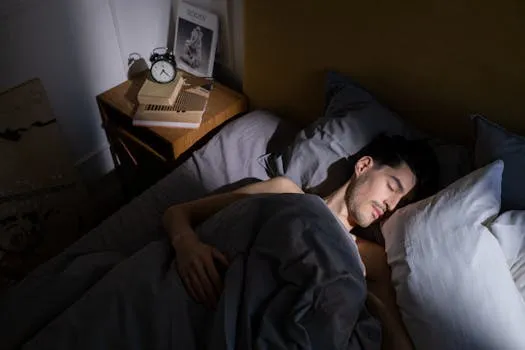
The reasons for under-diagnosis are systemic. Primary care visits are short. Sleepiness is normalized as a side effect of busy modern life. Snoring is treated as a benign annoyance rather than a clinical symptom. Sleep studies were historically expensive and required spending a night in a sleep lab — a barrier that home testing has substantially reduced. Women present with different symptom patterns than men (less snoring, more insomnia and fatigue) and are historically underscreened. And there remains a cultural tendency to minimize sleep disorders as less serious than “real” medical problems, despite the cardiovascular and metabolic data documenting their consequences.
The practical implication: unexplained fatigue, morning headaches, significant snoring, observed breathing pauses, or poor sleep quality despite adequate hours, without an evaluation for sleep apnea, mean there’s information not being acted on. The barriers to evaluation are lower than most people think, and the potential return is high.
Women and Sleep Apnea: The Diagnostic Gap
The conventional image of sleep apnea — a heavy-set middle-aged man who snores like a chainsaw and falls asleep mid-conversation — has been one of the more consequential diagnostic blind spots in sleep medicine. Women have OSA at roughly half the prevalence of men, but they present differently, are screened less frequently, and are diagnosed on average years later than men with comparable disease severity.
Female OSA presentation is more likely to involve insomnia, fatigue, morning headaches, depression, and anxiety than the classic male presentation of loud snoring and excessive daytime sleepiness. Women with OSA are more likely to have hypopneas (partial obstruction) rather than frank apneas (complete cessation), which are less dramatic but equally pathological. The STOP-BANG questionnaire weights male sex and BMI heavily — criteria that shift scoring away from women who may have significant OSA at lower body weight with less prominent snoring.
Hormonal factors modulate OSA risk across women’s lifespan in ways that affect diagnostic awareness. Progesterone is a respiratory stimulant that provides some protection against upper airway collapse, one reason premenopausal women have lower OSA prevalence than age-matched men. At menopause, progesterone levels fall dramatically, and OSA prevalence in postmenopausal women rises to approach male rates. A woman who never snored and never worried about sleep apnea may develop significant OSA at menopause, particularly in the context of menopausal weight gain that further narrows the airway.
Pregnancy OSA deserves specific mention. The combination of weight gain, nasal congestion from increased blood volume, hormone changes, and the mechanical effect of the uterus on diaphragm excursion creates substantially elevated OSA risk, particularly in the third trimester. Pregnancy OSA is associated with hypertensive disorders of pregnancy (preeclampsia), gestational diabetes, and preterm birth — making screening and treatment clinically important. A 2014 study in the American Journal of Obstetrics and Gynecology found that OSA was associated with significantly increased odds of preeclampsia, gestational diabetes, and other adverse pregnancy outcomes. Standard OSA screening tools are inadequate for pregnancy; any pregnant woman with snoring, witnessed apneas, or significant fatigue disproportionate to pregnancy norms deserves evaluation.
For women at perimenopause and menopause, routine OSA screening should arguably be standard practice. The intersection of worsening sleep quality from hormonal changes, increased OSA vulnerability from progesterone decline, and potential weight gain creating airway narrowing means the menopausal transition is a high-risk window for OSA emergence that often goes undetected because symptoms get attributed to “normal menopause.”
OSA, Cognitive Decline, and the Long Game
The connection between untreated sleep apnea and cognitive decline is emerging as one of the more important stories in dementia prevention research. The mechanism is increasingly well-understood, and it has urgent implications for anyone with untreated moderate-to-severe OSA.
The glymphatic system — the brain’s waste-clearance mechanism that operates during deep sleep — requires sustained, uninterrupted slow-wave sleep to function effectively. Every apnea event that fragments deep sleep is an interruption in the clearance cycle. In severe OSA, hundreds of these interruptions per night mean the glymphatic system is repeatedly rebooting rather than completing its clearance work. Over months and years, this impaired clearance allows metabolic waste products, including amyloid-beta and tau proteins, to accumulate in brain tissue.
The 2015 Nature Communications study by Ricardo Osorio found that sleep apnea was associated with earlier amyloid accumulation in the brain — by nearly five years on average compared to age-matched controls without OSA. This finding has been replicated and extended in subsequent research. Whether treating OSA reverses amyloid accumulation (rather than just slowing it) is an active research question. Several ongoing trials are examining whether CPAP treatment affects cognitive trajectory in older adults with both OSA and mild cognitive impairment.
The practical implication is straightforward: middle age, risk factors for OSA, and a family history of Alzheimer’s or dementia together mean the sleep apnea evaluation is not optional. The window for protective intervention likely exists, but it is not infinite. The treatment decision that felt like it could wait indefinitely has a timeframe attached to it now. Get evaluated. If it’s there, treat it — not just for cardiovascular risk and daytime function, but for the brain’s long-term health.
Paul’s Outcome
Paul was prescribed CPAP. He quit after three weeks because the mask gave him claustrophobia and he couldn’t tolerate the pressure. He went back to his sleep specialist, who tried an APAP unit with a nasal pillow mask instead of the full-face mask. Better. Then he added a chinstrap to stop mouth breathing that was causing air leaks. Better still. He also, prodded by his doctor’s directness about his 47-pound excess weight, started working with a nutritionist and a trainer.
Eighteen months later, Paul had lost 31 pounds. His repeat sleep study showed an AHI of 9 — mild OSA, down from severe. He still used CPAP but now with equipment that actually fit his face and settings that didn’t feel like trying to exhale against a wall. His wife moved back into the bedroom. His blood pressure, which had required medication for three years, came down enough that his cardiologist reduced his dose. He stopped falling asleep during afternoon meetings.
Paul’s story isn’t unusual. It’s unremarkable in the best possible way. Treat the apnea, lose the weight, and the body does what bodies are designed to do when they’re actually allowed to sleep. The tragedy is the fifteen years before the sleep study. The diagnosis came from an ultimatum. It could have come from awareness.
For the complete sleep optimization framework, see the Sleep Optimization Protocol. For broader health and recovery strategies, visit the Health hub.
Sleep Apnea Signs Q&A
Can I have sleep apnea if I don’t snore?
Yes. Snoring is a common symptom but not a required one. Women with OSA in particular frequently present without significant snoring, experiencing instead fatigue, insomnia, morning headaches, and mood disturbances. Central sleep apnea often occurs without snoring. Excessive daytime sleepiness, unrefreshing sleep, waking with headaches, waking with gasping — the absence of snoring shouldn’t be reassuring on its own.
Is sleep apnea really a health risk, or is it mostly a quality-of-life issue?
It’s both, and both dimensions are serious. The quality-of-life impacts (daytime fatigue, cognitive impairment, relationship strain from snoring) are real and significant. The health risks — 3x cardiovascular risk, 2x diabetes risk, accelerated cognitive decline, association with amyloid accumulation — are also real and documented in large prospective cohort studies. Treating it as a mere inconvenience misrepresents the medical evidence.
What’s the minimum AHI that needs treatment?
Treatment recommendations depend on AHI in combination with symptoms and comorbidities, not AHI alone. An AHI of 10 with significant daytime impairment and hypertension is typically treated. An AHI of 10 with no symptoms and no comorbidities may be observed with behavioral modification. An AHI above 15 (moderate) with any significant symptoms or cardiovascular comorbidities is generally treated. Above 30 (severe), treatment is almost universally recommended regardless of symptoms.
I tried CPAP and couldn’t tolerate it. Is that it?
Not even close. CPAP failure is common and treatable in many cases. Common failure modes include wrong mask type (full-face when nasal or nasal pillow would be tolerated), pressure settings too high or too low for comfort (APAP adjusts automatically), aerophagia from excess pressure, dry airway from lack of humidification, and inadequate desensitization period. Working with a sleep medicine specialist specifically focused on CPAP troubleshooting, rather than just being handed equipment and sent home, substantially improves adherence rates. If CPAP truly fails after proper troubleshooting, oral appliances, myofunctional therapy, and positional therapy are legitimate second-line options.
Will losing weight cure my sleep apnea?
Possibly, depending on severity and anatomy. For overweight and obese patients, significant weight loss (10-15% or more of body weight) produces clinically meaningful AHI reduction and in some cases complete remission. It’s the most powerful non-device intervention available. However, OSA in normal-weight individuals has a stronger anatomical component (jaw structure, soft palate anatomy) that is less responsive to weight change. And even with significant weight loss, some patients have residual OSA requiring treatment. Track with repeat sleep study after meaningful weight loss to determine residual severity.
My partner says I stop breathing in my sleep. How seriously should I take this?
Very seriously. Observed apneas are the most specific symptom of OSA — a partner watching you stop breathing during sleep means the likelihood of significant OSA is high. In the Wisconsin Cohort data, witnessed apneas were among the strongest predictors of clinically significant OSA. Get a sleep study. This is not an “eventually” recommendation.
Can children have sleep apnea?
Yes, and it’s underdiagnosed in children as well. Pediatric OSA often presents differently than adult OSA — symptoms include mouth breathing, restless sleep, behavioral problems, learning difficulties, and bed-wetting rather than primarily snoring and daytime sleepiness. Enlarged tonsils and adenoids are the most common anatomical cause in children. Unlike in adults, tonsillectomy and adenoidectomy are often curative in children with OSA driven by tonsillar hypertrophy. Early diagnosis and treatment matter because untreated pediatric OSA is associated with neurodevelopmental consequences including impaired attention, behavioral regulation, and academic performance.
The Practical Framework: Applying Sleep Apnea Signs Risks In Real Life
References
Editorial StandardsCorrectionsMedical DisclaimerAbout Our ContentAffiliate DisclosureSite Map