
Sarah was forty-one and exhausted in a way sleep didn’t fix. She’d been gaining weight despite not changing her diet. Her hair was coming out in the shower in amounts that alarmed her. She was cold in rooms where other people were comfortable. Constipated, foggy, and depressed in a muted, gray-curtain way that wasn’t dramatic enough to feel like “real” depression but was relentlessly, quietly present anyway. She’d had her thyroid tested twice. Both times her doctor looked at the TSH result — 3.1 mIU/L the first time, 2.9 the second — and said the same thing: “Your thyroid is completely normal.” What her doctor hadn’t ordered was a free T3, a free T4, a reverse T3, or a set of thyroid antibodies. What they’d been doing, in effect, was checking the thermostat and declaring the heating system fine without ever looking at whether heat was actually being produced and reaching the rooms. A complete thyroid panel, when she finally got one, told a more complicated and far more treatable story.
Why TSH Alone Is Insufficient
Thyroid-stimulating hormone is a pituitary hormone — produced by the pituitary gland, not the thyroid gland itself. TSH tells the thyroid to produce more thyroid hormone. Measuring TSH tells you what the pituitary is signaling; it does not tell you whether the thyroid is responding appropriately, whether the hormone being produced is being converted to its active form, whether that active form is reaching tissues, or whether autoimmune processes are damaging the thyroid even while current function still looks adequate.
A 2012 review by Biondi and Cooper in the New England Journal of Medicine synthesized the growing evidence that TSH alone misses substantial thyroid dysfunction — particularly in patients with subclinical hypothyroidism (TSH above optimal but below the standard “abnormal” threshold), conversion problems (normal TSH and T4 but impaired T4-to-T3 conversion), and early Hashimoto’s thyroiditis (antibody-positive but not yet affecting TSH). They estimated that TSH-only testing misses clinically significant thyroid dysfunction in a substantial minority of symptomatic patients.

Point 1 — Hypothalamus/Pituitary: Sends the TSH signal. Secondary hypothyroidism involves low TSH despite low thyroid hormones — the pituitary isn’t signaling correctly. TSH-only testing would miss this entirely (low TSH looks like hyperthyroidism, not hypothyroidism).
Point 2 — Thyroid Gland: Produces T4 in response to TSH. Autoimmune destruction (Hashimoto’s) may be active well before T4 production is significantly impaired. Antibodies can be positive for years before TSH ever moves.
Point 3 — Peripheral Conversion: T4 is converted to active T3 in the liver, gut, and other peripheral tissues. This conversion can be impaired by chronic stress (elevated cortisol), caloric restriction, gut dysfunction, or nutritional deficiencies (selenium, zinc, iodine all cofactor T4-to-T3 conversion). Normal TSH and normal T4 but low free T3 — TSH misses this entirely.
Point 4 — Receptor Level: Thyroid hormone resistance at the receptor level means adequate circulating T3 isn’t producing the expected cellular response. TSH and T3 can both look fine while cells remain functionally hypothyroid.
Testing TSH alone for thyroid function is like testing your car by checking the gas pedal position without looking at the fuel gauge, injection system, or engine output. The pedal position tells you one thing. It doesn’t tell you whether the car is actually running well.
TSH Optimal Range: The Evidence for 1-2 mIU/L
The standard TSH reference range of 0.5-4.5 mIU/L was derived statistically from population distributions, with the upper limit representing the 97.5th percentile of a “healthy” reference population. The problem — as with lab ranges more broadly — is that this population wasn’t actually healthy by functional criteria. A significant proportion of people with TSH in the 2.5-4.5 range have subclinical hypothyroid symptoms and elevated cardiovascular risk markers.
The research evidence for a tighter optimal range:
Rodondi et al. (2010, JAMA) — the largest systematic review and meta-analysis of subclinical hypothyroidism at the time — found that TSH above 10 mIU/L significantly elevated cardiovascular events, and that even TSH in the 4.5-10 range (still “normal” by some labs’ criteria) was associated with elevated cardiovascular risk in certain populations. Multiple subsequent studies have extended concern down to TSH above 2.5-3.0.
A comprehensive population study by Åsvold et al. (2012, published in JAMA Internal Medicine) examined the relationship between TSH and total cholesterol, LDL, and triglycerides in over 30,000 subjects. They found a linear association between TSH and cardiovascular risk markers — as TSH rose within the “normal” range, cardiovascular risk markers worsened, with no threshold effect anywhere. There was no safe “normal” TSH. Risk increased continuously with TSH level, full stop.
The American Thyroid Association’s guidelines have been progressively narrowing the target TSH range for treatment, with a 2019 update acknowledging that TSH of 1.0-2.5 mIU/L may be more appropriate for some patients than the broader reference range. The functional medicine consensus, based on this research and clinical experience with symptom correlation, has settled on 1.0-2.0 mIU/L as the zone where most patients feel and function best.
Sarah’s TSH of 2.9-3.1 was technically “normal” by every standard lab criterion. It sat at the border of where the research suggests symptoms begin to emerge and cardiovascular risk begins to climb. Her clinical picture — every symptom consistent with hypothyroidism — told the same story. The TSH alone had been ambiguous enough to wave off. The complete panel wouldn’t be.
Free T3: The Most Important Marker That Gets Ignored
Triiodothyronine (T3) is the biologically active thyroid hormone. While the thyroid produces some T3 directly, roughly 80% of circulating T3 comes from peripheral conversion of thyroxine (T4) through the action of deiodinase enzymes. T3 is the form that actually enters cells, binds thyroid receptors, and drives metabolic rate, body temperature, heart rate, protein synthesis, and dozens of other functions that define thyroid health.
“Free” T3 refers to the unbound fraction of T3 in circulation — the fraction actually available to enter cells. The majority of circulating T3 is bound to carrier proteins (primarily thyroxine-binding globulin) and is biologically inactive. Free T3 is the number that matters.
Standard lab reference ranges for free T3 typically span 2.0-4.4 pg/mL. The research on symptom burden and metabolic function suggests optimal free T3 sits at 3.0-4.0 pg/mL — the upper-middle portion of the reference range. People with free T3 at 2.0-2.5 pg/mL may be technically “within range” while experiencing significant hypothyroid symptoms, because their cellular T3 delivery sits at the low end of functional.
The conversion problem: in chronic stress states (elevated cortisol), severe caloric restriction (crash diets), significant gut inflammation, or after major illness, the deiodinase enzymes that convert T4 to T3 get downregulated. The body preferentially converts T4 to reverse T3 (an inactive isomer) rather than active T3 — an adaptive mechanism built to reduce metabolic rate during periods of stress or resource scarcity. The result is normal TSH, normal T4, low free T3, high reverse T3, and every symptom of hypothyroidism despite what looks, by TSH criteria, like a “normal” thyroid.
This conversion problem is completely invisible to TSH testing. It requires measuring free T3 and reverse T3 simultaneously to catch.
Free T4: The Precursor Marker
Thyroxine (T4) is the primary hormone produced directly by the thyroid gland — roughly 90% of thyroid output is T4, with about 10% being direct T3 production. T4 itself has minimal biological activity; it functions primarily as a precursor to T3. “Free” T4 is the unbound, bioavailable fraction.
Measuring free T4 alongside free T3 accomplishes two important things: it confirms the thyroid is producing adequate T4 (a production problem and a conversion problem look different on these tests), and it establishes T4-to-T3 conversion efficiency when compared against the free T3 result.
If free T4 is low with elevated TSH — the pituitary is shouting at a thyroid that isn’t producing enough T4. Classic primary hypothyroidism. If free T4 is normal or high but free T3 is low — T4 is being produced but not efficiently converted to the active form. A conversion problem, and one that standard thyroid treatment (typically T4-only medications like Levothyroxine) often doesn’t touch.
Optimal free T4: 1.1-1.7 ng/dL (the upper-middle portion of the typical 0.8-1.8 ng/dL reference range). Low-normal free T4 combined with low-normal free T3 despite normal TSH is a pattern suggesting both suboptimal thyroid output and impaired conversion — a finding pure TSH testing would completely miss.
Reverse T3: The Invisible Braking System

Reverse T3 is produced by the same deiodinase enzymes that produce active T3, but under stress conditions the equilibrium shifts toward rT3 production. Part of the “euthyroid sick syndrome,” or “low T3 syndrome” — a conserved adaptive response in mammals to severe physical stress (illness, fasting, surgery, trauma) that reduces metabolic rate to preserve energy.
The problem is that in modern contexts, non-physical stressors — chronic psychological stress, sleep deprivation, extreme dieting, and some medications — can activate the same rT3-upregulating pathway without the actual survival threat the adaptation was built for. The result is chronically elevated rT3 creating functional hypothyroidism in people who aren’t physically ill at all.
Standard reference range for rT3 is typically 10-24 ng/dL. The more relevant metric is the free T3 to reverse T3 ratio: divide the free T3 (in pg/mL) by the reverse T3 (in ng/dL) and multiply by 100. Optimal ratio is above 20. Below 20 suggests rT3 is competing meaningfully with active T3 for receptor binding and producing functional hypothyroid effects.
The rT3 story explains why some people gain weight dramatically on very low-calorie diets — the body interprets caloric restriction as a threat, upregulates rT3, reduces active T3, and effectively down-regulates metabolism to conserve resources. Standard thyroid tests remain “normal” throughout this entire process.
TPO and TG Antibodies: Early Warning for Hashimoto’s
Hashimoto’s thyroiditis is the most common cause of hypothyroidism in developed countries — an autoimmune condition where the immune system attacks thyroid tissue, gradually destroying function over years to decades. The distinguishing feature of Hashimoto’s is the presence of antibodies against thyroid-specific proteins.
TPO antibodies (anti-thyroid peroxidase): Antibodies against the enzyme responsible for thyroid hormone synthesis. Present in 95% of Hashimoto’s cases. TPO antibody positivity can precede thyroid function abnormalities by years — the immune attack is underway long before TSH changes. A person with positive TPO antibodies and high-normal TSH (2.5-4.5) is very likely to progress to overt hypothyroidism and should be monitored closely.
TG antibodies (anti-thyroglobulin): Antibodies against thyroglobulin, the protein matrix in which thyroid hormone is stored. Present in about 60-80% of Hashimoto’s cases. Some people have positive TG antibodies without positive TPO, which is why both are necessary for a complete autoimmune screen.
Why does early detection of Hashimoto’s matter beyond just monitoring function? A few reasons.
First, dietary interventions — particularly gluten elimination — have some evidence for reducing TPO antibody levels and slowing autoimmune progression in Hashimoto’s patients. The molecular mimicry hypothesis (gliadin proteins in wheat share structural similarity with thyroid tissue, potentially triggering or amplifying autoimmune responses) has research support in a subset of Hashimoto’s patients. Knowing there’s Hashimoto’s present changes the dietary management approach entirely.
Second, selenium supplementation (200mcg daily) has been shown in multiple randomized trials to reduce TPO antibody levels in Hashimoto’s patients. The effect is meaningful — a meta-analysis by Wichman et al. (2016) found that selenium supplementation reduced TPO antibody levels by roughly 30% versus placebo. An available, inexpensive intervention — but only relevant once someone knows the antibodies are elevated.
Third, Hashimoto’s is an autoimmune condition, which means identifying it changes the entire health surveillance strategy going forward — other autoimmune conditions (celiac disease, type 1 diabetes, other thyroid conditions) turn up more often in people with one autoimmune condition already, and proactive screening starts to make sense.
The Complete Thyroid Interpretation Guide Framework
Here’s how to interpret a complete thyroid panel systematically. For each combination of results, the clinical picture and likely mechanisms differ:
Pattern 1 — Subclinical Hypothyroidism: TSH above 2.5 mIU/L, free T4 low-normal, free T3 low-normal, antibodies negative. Suggests primary underactivity without autoimmune cause. May respond to lifestyle interventions (iodine and selenium optimization, stress reduction) or may require low-dose thyroid hormone support if symptomatic.
Pattern 2 — Hashimoto’s (Early): TSH borderline (2.0-4.0), free T4 and T3 may be normal, TPO and/or TG antibodies positive. Autoimmune process underway, function not yet significantly impaired. Intervention: dietary approaches (gluten evaluation, selenium supplementation), close monitoring, addressing other autoimmune triggers.
Pattern 3 — Conversion Problem: TSH normal or low-normal, free T4 normal, free T3 low, reverse T3 elevated, rT3/T3 ratio below 20. T4 production is adequate, conversion to active T3 is impaired. Common in chronic stress, caloric restriction, illness. Intervention: address the root cause (stress, diet, sleep), optimize conversion cofactors (selenium, zinc, iron — all required for deiodinase function).
Pattern 4 — Overt Hypothyroidism: TSH above 4.5 (or significantly above optimal), free T4 below range, free T3 below range. Classic primary hypothyroidism requiring thyroid hormone replacement. The only pattern TSH-alone testing reliably catches.
Pattern 5 — Secondary/Central Hypothyroidism: TSH low or normal, free T4 low, free T3 low. Pituitary or hypothalamic dysfunction rather than thyroid gland failure. TSH-only testing would typically misclassify this as hyperthyroid (low TSH) or normal. Requires endocrinology evaluation.
Pattern 6 — Subclinical Hyperthyroidism: TSH below 0.5 mIU/L, free T4 and T3 normal or high-normal. May stem from excessive thyroid hormone medication, a toxic nodule, or early Graves’ disease. Associated with atrial fibrillation risk and bone density loss over time.
Nutrients That Support Thyroid Function
Several nutritional deficiencies directly impair thyroid function and can produce or worsen hypothyroid patterns. Optimizing these is appropriate before or alongside medical treatment:
Iodine: The essential building block of thyroid hormones — T4 contains four iodine atoms, T3 contains three. Iodine deficiency is the world’s most common cause of hypothyroidism and the primary reason iodine was added to salt in the 20th century. In the US, iodine sufficiency should be evaluated before supplementing — excessive iodine in Hashimoto’s patients can actually worsen autoimmune activity. Test through urinary iodine if concerned.
Selenium: A cofactor for the deiodinase enzymes that convert T4 to T3, and for the selenoprotein P that protects the thyroid from oxidative damage. Selenium deficiency directly impairs T3 production. Brazil nuts (2 per day provides adequate selenium), or 100-200mcg supplemental selenomethionine. Particularly important for Hashimoto’s patients given the antibody-reduction evidence.
Zinc: Required for thyroid hormone receptor function and T4-to-T3 conversion. Zinc deficiency impairs cellular response to thyroid hormones even when serum levels look adequate. 15-30mg supplemental zinc or high dietary intake from meat, shellfish, and seeds.
Iron: Thyroid peroxidase — the enzyme that synthesizes thyroid hormones — is an iron-dependent enzyme. Iron deficiency impairs thyroid hormone production and can cause TSH elevation that reverses with iron repletion alone. If ferritin is below 40 ng/mL and TSH is elevated, iron status should be addressed before or alongside thyroid treatment.
Working the Medical System for Thyroid Care
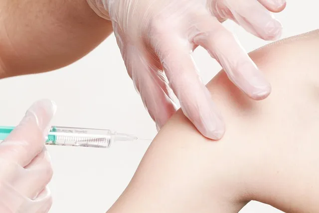
Direct-to-consumer thyroid testing is available through multiple services (Ulta Lab Tests, Everly Well, LabCorp Direct) and allows ordering the complete panel — TSH, fT3, fT4, rT3, TPO and TG antibodies — without a physician order, for roughly $100-200. This gets the data on hand before the clinical conversation even happens.
When choosing a physician for thyroid evaluation, specifically look for practitioners who order the full panel, use optimal ranges (TSH 1-2 mIU/L, not 0.5-4.5), are comfortable prescribing combination T4/T3 therapy when indicated (some hypothyroid patients do better on T4+T3 or natural desiccated thyroid than on T4-only treatment), and treat symptoms alongside numbers rather than instead of them. A physician who dismisses symptoms because TSH is “normal,” without ever ordering a complete panel, is the wrong physician for this kind of evaluation.
Sarah’s complete thyroid panel showed: TSH 3.1 (technically normal, functionally borderline), free T4 0.9 ng/dL (low normal), free T3 2.2 pg/mL (below optimal 3.0-4.0), reverse T3 28 ng/dL (elevated), and TPO antibodies 450 IU/mL (significantly elevated — normal is below 35). She had Hashimoto’s thyroiditis with active autoimmune attack, impaired T4-to-T3 conversion, and a TSH that had been masking the full picture the entire time. The treatment path — addressing autoimmune triggers, optimizing conversion cofactors, targeted thyroid hormone support — was finally clear. None of it had been visible through a single TSH number. All of it had been there the whole time, waiting for the right questions to be asked.
Reader Questions About Thyroid Panel Optimal
- My TSH is 3.2 and my doctor says it’s normal. Should I get a full panel?
Yes, particularly with symptoms consistent with hypothyroidism (fatigue, weight gain, hair loss, cold intolerance, constipation, brain fog, depression). TSH of 3.2 sits in the range where the research shows elevated symptom burden and cardiovascular risk markers, and where Hashimoto’s and conversion problems are commonly found. The full panel — fT3, fT4, rT3, TPO and TG antibodies — costs roughly $100-150 out of pocket through direct-to-consumer labs and provides information that completely changes the clinical picture. - What’s the difference between Hashimoto’s and general hypothyroidism?
General hypothyroidism is the result — insufficient thyroid hormone. Hashimoto’s thyroiditis is the most common cause: an autoimmune condition where antibodies attack thyroid tissue, gradually destroying function. The distinction matters because Hashimoto’s is an autoimmune condition with specific management approaches (dietary modification, selenium supplementation, monitoring for other autoimmune conditions, gluten evaluation) beyond simply replacing thyroid hormone. A standard hypothyroidism diagnosis says the thermostat is off; a Hashimoto’s diagnosis says why, and opens different treatment options entirely. - Can I improve thyroid function without medication?
Depending on the pattern, yes — in some cases significantly. Conversion problems (low fT3, high rT3 despite normal TSH) often respond to addressing root causes: reducing chronic stress, improving sleep, ensuring caloric adequacy, optimizing selenium and zinc status. Early Hashimoto’s may respond to dietary approaches and selenium supplementation in terms of antibody reduction and slowing progression. Established overt hypothyroidism (low fT4, elevated TSH) typically requires thyroid hormone replacement — lifestyle optimization supports but doesn’t replace pharmaceutical treatment at that stage. - Is Levothyroxine (T4-only medication) adequate for everyone?
Not for everyone. Levothyroxine provides T4, which must be converted to active T3 in peripheral tissues. Most people convert adequately. But individuals with impaired deiodinase function — due to genetic variants in DIO2 (the conversion enzyme gene), chronic stress, nutritional deficiencies, or gut dysfunction — don’t convert T4 to T3 efficiently and may remain symptomatic on T4-only treatment despite “normal” TSH. These patients often do better on combination T4/T3 therapy (Levothyroxine + Liothyronine) or natural desiccated thyroid (NDT), which contains both hormones. Measuring free T3 while on T4-only treatment reveals whether conversion is actually adequate. - How does selenium help with Hashimoto’s?
Selenium is a cofactor in selenoprotein P, which protects the thyroid from oxidative damage generated during thyroid hormone synthesis. Thyroid tissue has very high selenium requirements relative to its size. In Hashimoto’s, autoimmune-driven inflammation generates oxidative stress that damages thyroid tissue; adequate selenium supports the antioxidant systems protecting against this damage. Multiple randomized trials (summarized in the Wichman 2016 meta-analysis) have found that 200mcg daily selenomethionine reduces TPO antibody levels by roughly 30% and may slow disease progression in Hashimoto’s patients. One of the few nutritional interventions in Hashimoto’s with consistent trial evidence behind it. - What does elevated reverse T3 mean and how do I fix it?
Elevated reverse T3 (rT3) indicates the body is preferentially converting T4 to the inactive isomer rather than active T3 — effectively putting the brakes on thyroid function at the cellular level. This typically occurs in response to physiological stress signals: chronic psychological stress (elevated cortisol), severe caloric restriction, prolonged illness, or sleep deprivation. The fix is addressing the root cause — reduce chronic stress (sleep, meditation, workload management), ensure adequate caloric and nutritional intake, address sleep quality, and optimize selenium and zinc for conversion enzyme support. If rT3 remains elevated after all that, formal thyroid evaluation with a functional medicine physician is appropriate. - Should I take iodine supplements for thyroid health?
Not without testing first. While iodine deficiency impairs thyroid hormone synthesis, iodine excess — particularly in people with Hashimoto’s — can actually worsen autoimmune thyroid disease by triggering more aggressive immune responses against thyroid tissue. A well-documented clinical problem. Anyone living in a developed country, using iodized salt, eating seafood and dairy, is unlikely to be iodine deficient. Test urinary iodine before supplementing. And with Hashimoto’s specifically, iodine supplementation should be discussed carefully with a knowledgeable physician before proceeding.
The thyroid system is too complex to evaluate with a single marker, and the consequences of missing dysfunction are too significant to accept inadequate testing as the standard. The Complete Thyroid Interpretation Guide — TSH within optimal range, free T3 in the active zone, free T4 confirming production adequacy, reverse T3 confirming efficient conversion, and antibodies confirming absence of autoimmune activity — gives a complete picture that TSH alone categorically cannot provide. Getting this panel done once, especially with symptoms consistent with thyroid dysfunction, may be the highest-value diagnostic step available in preventive health. Sarah’s story is common. The information to change it is accessible. The barrier is usually just knowing which questions to ask.
Related reading: Blood Test Results Explained: Optimal vs Normal | Annual Blood Panel: 30 Markers Every Man Needs
How to Talk to Your Doctor About a Complete Panel
One of the most common barriers to getting a complete thyroid workup is the physician conversation itself. Many patients leave appointments without the tests they need because they didn’t know how to ask, or because the physician’s first response was dismissive. Having a prepared framework makes this conversation more productive.
Start by framing the request around symptoms, not self-diagnosis. “I’ve been experiencing persistent fatigue, hair thinning, cold intolerance, and cognitive sluggishness for several months, and I’d like a complete thyroid panel that includes free T3, free T4, reverse T3, and thyroid antibodies — not just TSH.” This positions the patient as someone tracking their own health, not someone challenging clinical authority. If the physician resists, a specific follow-up question helps: “Given that my TSH is in the 2.5-3.5 range and I have multiple symptoms consistent with thyroid dysfunction, what is your clinical rationale for not measuring free T3 and antibodies?” A physician who can’t answer that concisely is likely defaulting to habit rather than clinical reasoning.
If a physician refuses entirely, the direct-to-consumer route is a practical bypass. Ulta Lab Tests, Walk-In Lab, and LabCorp Direct all offer the complete thyroid panel without a physician order for $80-150 in most US states. Results can be brought in hand for a more informed conversation. Alternatively, a second opinion from an integrative physician, a naturopathic doctor with thyroid specialization, or a functional medicine practitioner — clinicians who routinely order the full panel and interpret it using optimal rather than standard ranges — is worth seeking out.
Documenting symptoms before the appointment helps too. A dated symptom log — “fatigue started in month X, hair loss in month Y, weight gain of Z pounds over W months” — provides the clinical narrative that makes the case more compelling and harder to dismiss. Symptoms with documented onset and progression are harder to wave off as “stress” or “aging” than a vague complaint squeezed into a 10-minute appointment.
Thyroid Health After 40: What Changes and Why
Thyroid function changes meaningfully with age, and these changes are worth understanding for anyone doing ongoing monitoring. Several specific patterns become more common after 40 in both men and women.
Conversion efficiency declines with age. The deiodinase enzymes responsible for converting T4 to active T3 become less efficient in older adults, meaning that even with adequate T4 production and normal TSH, free T3 levels tend to run lower in older age groups. A biological reality, not a failure — the body is down-regulating metabolic rate as part of the aging process. But it also means the “normal” range for free T3 in a 60-year-old who wants to maintain energy and metabolic function may require active management that a 30-year-old simply doesn’t need.
Hashimoto’s prevalence increases steadily with age, with peak incidence in women between 45 and 65. The post-menopausal hormonal shift, which changes immune regulation, appears to increase susceptibility to autoimmune thyroid attack. Any woman in perimenopause or beyond who hasn’t had thyroid antibodies tested is operating with incomplete information about one of the most common health conditions affecting her demographic.
For men, testosterone decline and thyroid function interact bidirectionally. Low testosterone reduces T4-to-T3 conversion efficiency. Low thyroid function reduces testosterone production. Men experiencing symptoms of both low testosterone and low thyroid (fatigue, low libido, weight gain, cognitive fog, decreased motivation) often have both conditions simultaneously, and treating only one frequently produces incomplete recovery. Evaluating both the complete thyroid panel and total/free testosterone together is standard practice in functional medicine for symptomatic men over 40.
Monitoring frequency after 40 should increase if any panel marker is suboptimal. Annual testing of the full panel — TSH, free T3, free T4, antibodies — is reasonable for anyone with previous borderline results, positive antibodies, or ongoing symptoms. Two-year monitoring is appropriate for those with completely normal results and no symptoms. The thyroid is a dynamic system that shifts over years; a single clean panel doesn’t guarantee clean panels indefinitely.
The Thyroid-Gut Connection: An Often-Missed Link
The relationship between gut health and thyroid function is bidirectional, clinically significant, and systematically neglected in conventional thyroid management. Understanding it adds a practical intervention lever that most people with thyroid dysfunction have never been offered.
Roughly 20% of T4-to-T3 conversion occurs in the gut — specifically through the action of gut bacteria producing deiodinase-like enzymes and through direct gut epithelial conversion. Gut dysbiosis (imbalanced gut microbiome), intestinal permeability (“leaky gut”), and small intestinal bacterial overgrowth (SIBO) all impair this conversion pathway. Research has consistently found patients with Hashimoto’s showing higher rates of intestinal permeability and altered gut microbiome composition compared to healthy controls. The relationship isn’t incidental.
The immune connection: roughly 70% of the immune system resides in the gut-associated lymphoid tissue (GALT). Hashimoto’s is an immune system dysregulation — the same gut-based immune dysfunction that drives intestinal permeability and autoimmune GI conditions also appears to amplify thyroid autoimmunity. Which is why Hashimoto’s co-occurs with celiac disease at rates far above chance — both conditions involve immune reactivity to antigens crossing an abnormally permeable gut barrier.
The practical implications: for anyone with Hashimoto’s or elevated thyroid antibodies, a gut-focused assessment and intervention is appropriate alongside thyroid-specific treatment. Evaluating for celiac disease (full celiac panel: tissue transglutaminase IgA, total IgA, deamidated gliadin peptide) before eliminating gluten is more informative than blindly going gluten-free. If celiac is excluded, a 3-month trial of gluten elimination still has meaningful evidence for reducing TPO antibodies in a significant subset of Hashimoto’s patients. A comprehensive stool analysis, SIBO breath testing, and gut-support protocols (digestive enzymes, probiotics, zinc carnosine for gut barrier integrity) are the first-line gut interventions for a thyroid-autoimmune presentation.
Sarah’s story above is about a complete thyroid panel. But for many people with Hashimoto’s, the complete protocol also includes gut repair — because the thyroid won’t fully stabilize while the gut-based immune dysregulation driving antibody production goes unaddressed. The thyroid panel is the diagnostic tool. The gut is one of the root causes.
The Thyroid-Weight Connection: Why Standard Advice Fails Hypothyroid Patients
Weight gain is one of the most common and frustrating symptoms of hypothyroidism, and one of the most mismanaged. Standard dietary advice — eat less, move more — gets dispensed reflexively at every appointment, without accounting for the specific metabolic changes thyroid dysfunction produces. Understanding the physiology of thyroid-driven weight gain reveals why caloric restriction alone rarely works, and sometimes makes the situation worse.
Thyroid hormone — specifically T3 — is the primary regulator of resting metabolic rate. T3 determines how many calories the body burns at rest to maintain basic physiological functions: maintaining cell membrane sodium-potassium gradients, producing heat, powering organ function. When T3 is low — whether from inadequate thyroid production, impaired conversion, or receptor resistance — resting metabolic rate decreases. Research by Reinehr (2010) and others has consistently shown that even subclinical hypothyroidism (TSH above 2.5 with normal or low-normal T3) is associated with measurably reduced resting energy expenditure. Not psychological. Not a matter of willpower. The metabolic engine is running at a lower speed because the thyroid throttle is partially closed.
The caloric restriction trap: when a hypothyroid person with reduced metabolic rate restricts calories significantly to lose weight, the body interprets the caloric deficit as a threat and activates the stress-adaptive conversion pathway — upregulating reverse T3 production and downregulating active T3 further still. Metabolic rate decreases in response to the caloric restriction, eliminating or reversing the very deficit that was supposed to produce weight loss. This is the mechanism behind the extremely common clinical experience of hypothyroid patients who eat very little, exercise moderately, and keep gaining weight anyway — they’re in a physiological state where the normal thermodynamic equation (calories in, calories out) has been disrupted at the metabolic rate denominator.
The approach that works for hypothyroid weight management is therefore different from standard weight loss advice. It begins with optimizing thyroid function — particularly free T3, the metabolic rate regulator. Until T3 sits in the optimal range (3.0-4.0 pg/mL), metabolic rate is suppressed and weight loss attempts are working against the physiology rather than with it. Aggressive caloric restriction during untreated or undertreated hypothyroidism worsens the conversion problem. Moderate, sustainable caloric adjustment (a deficit of no more than 300-400 calories per day) combined with resistance training (which preserves lean mass and maintains the muscle component of resting metabolic rate) is the approach compatible with hypothyroid physiology. Protein prioritization (1.7-2.0g/kg body weight) prevents muscle loss during the caloric deficit and supports conversion enzymes that require adequate protein availability.
Once thyroid hormones are optimized — particularly once T3 is brought into the optimal range through treatment or conversion support — resting metabolic rate often increases meaningfully. Patients frustrated by years of weight resistance sometimes see spontaneous weight normalization as thyroid function improves, with modest dietary adjustments rather than aggressive restriction. The weight wasn’t a behavior problem. It was a hormone problem. That distinction matters for how it gets addressed.
FROM THE LIBRARY ›
References
Editorial StandardsCorrectionsMedical DisclaimerAbout Our ContentAffiliate DisclosureSite Map