

“You’ll want to follow up with your doctor about that.” Sandra went home and made the mistake of googling “thyroid nodule.” Within forty minutes she’d convinced herself she had thyroid cancer, was planning how to tell her children, and had already mentally fast-forwarded through surgery, radiation, and an uncertain future.
What she didn’t know — what the search results didn’t clearly communicate — was that thyroid nodules are extraordinarily common, discovered incidentally in roughly half of all adult necks examined with ultrasound, and that the overwhelming majority are completely benign and require nothing more than periodic monitoring. Her path from casual incidental finding to near-panic was a failure of medical communication that plays out thousands of times every day in clinics across the country.
Thyroid nodules are one of the most common clinical findings in modern medicine and simultaneously one of the most anxiety-provoking. The vast majority are benign. A small minority harbor malignancy. The art of thyroid nodule evaluation is precisely distinguishing between these groups — doing enough evaluation to find the cancers that need treatment while avoiding the cascade of unnecessary procedures, surgery, and anxiety that follows from over-investigating the majority of benign nodules.
Understanding this evaluation framework gives patients the context to work through a process that would otherwise feel frightening and opaque.
The Epidemiology: How Common Thyroid Nodules Actually Are
The first thing to understand about thyroid nodules is how extraordinarily prevalent they are — a fact that fundamentally reframes the anxiety most patients feel on receiving this diagnosis.
Palpable thyroid nodules — those detectable by physical examination — are found in approximately 4-7% of the general adult population in iodine-sufficient countries. But palpable nodules are the minority. High-resolution ultrasound, which can detect nodules smaller than 1 mm, finds thyroid nodules in 19-67% of randomly selected adults when examined by experienced sonographers. The wide range reflects differences in study populations, ultrasonography equipment quality, and the minimum size considered reportable.
Autopsy studies are particularly revealing. Post-mortem examination of thyroid glands in people who died of unrelated causes finds nodules in approximately 50% of specimens, many containing multiple nodules. The vast majority of these are benign colloid nodules, adenomas, or cysts — findings that were completely silent in life and would never have caused symptoms or disease had the person not happened to die of something else and have their thyroid examined.
The prevalence of thyroid nodules increases with age (uncommon in young adults, common in those over 60), with female sex (nodules occur 4 times more frequently in women than men), with history of head and neck radiation (dramatically increases nodule prevalence and malignancy risk), and with iodine deficiency (endemic goiter regions have higher nodule rates). Geographic variation exists: areas with high dietary iodine have different nodule characteristics than iodine-deficient regions.
Against this backdrop of enormous benign nodule prevalence, what’s the actual rate of thyroid cancer? Approximately 5-10% of nodules that undergo biopsy are found to contain thyroid cancer. However, that figure applies to nodules already selected for biopsy — a population enriched for higher-risk features. The true malignancy rate among all thyroid nodules detected by ultrasound is lower, estimated at approximately 3-5%.
And even among thyroid cancers, the majority are papillary thyroid carcinomas with indolent biology and excellent prognosis. What doctors mean by “thyroid cancer” and what the word “cancer” conjures by default are, in most cases, vastly different disease entities.
Ultrasound: The Foundation of Nodule Risk Stratification
Thyroid ultrasound is the foundation of nodule evaluation — more informative than physical examination, more accessible and lower-risk than biopsy, and capable of characterizing features that reliably predict malignancy risk. Understanding what radiologists and endocrinologists are looking for on ultrasound demystifies the evaluation process considerably.
Several validated risk stratification systems exist, the two most widely used being ACR TIRADS (Thyroid Imaging Reporting and Data System, from the American College of Radiology) and ATA (American Thyroid Association) guidelines. These systems score nodules based on specific ultrasound features and assign a risk category that guides biopsy recommendations.
The ultrasound features most strongly associated with malignancy include: solid composition (versus cystic or mixed); hypoechogenicity (the nodule appears darker than surrounding thyroid tissue, particularly “marked hypoechogenicity” — darker than the strap muscles of the neck); irregular or ill-defined margins; microcalcifications (tiny calcium deposits within the nodule, appearing as small bright spots on ultrasound); a taller-than-wide shape on transverse view; and evidence of extrathyroidal extension.
These features are derived from pathological correlations — tissue analysis after thyroid surgery identified which ultrasound characteristics corresponded to malignant versus benign histology at the population level.
The ACR TIRADS system assigns points for each suspicious feature and categorizes nodules as TR1 (benign), TR2 (not suspicious), TR3 (mildly suspicious), TR4 (moderately suspicious), or TR5 (highly suspicious). Each category has corresponding recommendations based on nodule size — what triggers biopsy in a TR5 nodule (2 cm) requires a larger size threshold for TR4 (2.5 cm) and larger still, or observation-only, for TR3.
Two nodule types are essentially never biopsied regardless of size: pure cysts (fluid-filled sacs with no solid component) are benign by definition and are simply monitored or drained if symptomatic. Spongiform nodules — appearing as aggregated tiny cysts with an almost “honeycomb” appearance — have a benign risk greater than 98%, and biopsy guidelines recommend observation rather than sampling.
The 2023 update to ACR TIRADS guidelines further refined the risk scoring, incorporating emerging data on vascularity patterns, elastography (tissue stiffness measurements), and nodule growth rate into more detailed risk assessment. High-quality thyroid ultrasound performed by experienced sonographers using modern equipment is a genuinely sophisticated diagnostic tool — not a screening test but a characterization test that, in expert hands, can narrow the malignancy probability to within a fairly tight range.
Thyroid Function Tests: What They Tell You About Nodules
Thyroid function tests — primarily TSH (thyroid-stimulating hormone) and, when indicated, free T4 and T3 — are an important but often misunderstood component of nodule evaluation. They don’t diagnose nodule malignancy, but they provide context that significantly affects management.
TSH level is the most important initial lab test when a thyroid nodule is discovered. A suppressed TSH (below normal range) suggests hyperthyroidism, which in the presence of a nodule raises the possibility of a “hot” or autonomous nodule — one producing thyroid hormone independently of TSH regulation. Hot nodules are overwhelmingly benign; autonomous functioning nodules rarely harbor thyroid cancer.
A low TSH in a nodule patient should prompt a thyroid radionuclide scan (radioactive iodine or technetium scan) to determine if the nodule is autonomous. If confirmed autonomous (“hot” on scan), biopsy is generally unnecessary.
Conversely, a normal or elevated TSH is more consistent with a non-functioning nodule — one that doesn’t produce thyroid hormone — and these are the nodules that undergo standard risk stratification and potential biopsy. Some studies have found that higher TSH levels within the normal range are associated with a slightly higher malignancy rate in nodules, though the association is modest and doesn’t independently change biopsy recommendations.
Thyroglobulin — a protein produced by thyroid cells — is not useful for evaluating new thyroid nodules. It’s elevated in virtually all thyroid conditions (benign and malignant) and lacks specificity. Calcitonin, however, has a specific role: it screens for medullary thyroid carcinoma (MTC), which accounts for approximately 5% of thyroid cancers and originates from parafollicular C cells. Some guidelines (particularly European) recommend routine calcitonin testing in all nodule patients; American guidelines are more selective.
Markedly elevated calcitonin strongly suggests MTC and changes the urgency of surgical evaluation.
Fine-Needle Aspiration Biopsy: What Happens and What It Tells You
- Bethesda I (Non-diagnostic/Unsatisfactory): Inadequate sample, insufficient cells for interpretation. Typically 1-4% malignancy risk, but requires repeat biopsy. Approximately 5-10% of FNA results.
- Bethesda II (Benign): Benign colloid nodule, cyst, or thyroiditis. Approximately 0-3% malignancy risk. No surgery needed; surveillance ultrasound recommended.
- Bethesda III (Atypia of Undetermined Significance/AUS): Architectural or cellular abnormality that doesn’t meet either benign or malignant criteria. Approximately 10-30% malignancy risk. Management varies — repeat FNA, molecular testing, or diagnostic lobectomy depending on clinical context.
- Bethesda IV (Follicular Neoplasm/Suspicious for Follicular Neoplasm): Follicular architecture raising concern for follicular adenoma vs. carcinoma — a distinction that cannot be made on FNA alone (requires surgical removal and examination of the capsule). Approximately 25-40% malignancy risk. Diagnostic lobectomy recommended.
- Bethesda V (Suspicious for Malignancy): Strong cytological concern without diagnostic certainty. Approximately 50-75% malignancy risk. Surgical resection recommended.
- Bethesda VI (Malignant): Diagnostic of thyroid carcinoma. Approximately 97-99% malignancy risk. Surgical resection required.
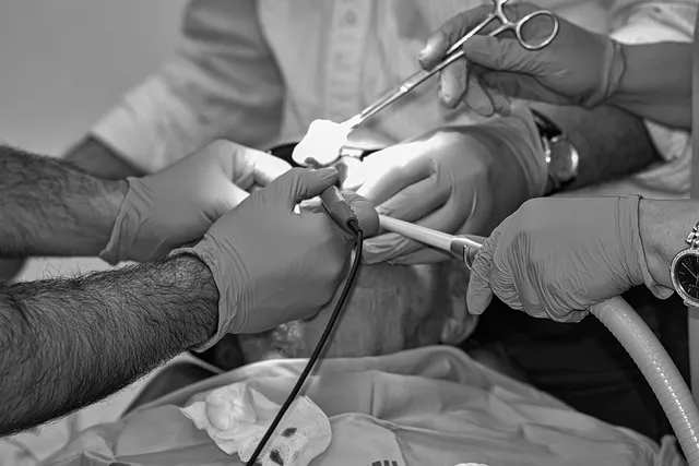
The procedure itself involves placing a thin needle (22-25 gauge — similar in diameter to an acupuncture needle) directly into the nodule under ultrasound guidance, aspirating cells from multiple passes through the nodule, and spreading the sample onto slides for cytological analysis. The procedure takes approximately 10-15 minutes in experienced hands, requires only topical or local anesthesia, and carries minimal risk — primarily minor discomfort and occasional small hematoma at the biopsy site.
Serious complications (infection, significant bleeding, nerve injury) are rare, occurring in less than 1% of procedures when performed under ultrasound guidance.
The results of FNA biopsy are classified using the Bethesda System for Reporting Thyroid Cytopathology — a six-category classification system with specific cancer risk estimates for each category:
Understanding these categories prevents a common and distressing experience: patients who receive a Bethesda III result are often told “we’re not sure — it might be cancer” and experience this as deeply alarming when, in reality, most Bethesda III nodules are benign and the uncertainty reflects cytological limitations, not high malignancy probability.
Molecular Testing: A Revolution in Indeterminate Nodule Management
The categories most challenging for management are Bethesda III and IV — the “indeterminate” categories where cytology cannot definitively classify the nodule. Historically, management of indeterminate nodules defaulted to diagnostic surgery in most cases, resulting in thousands of thyroid operations annually that revealed benign pathology — unnecessary surgery with real costs and risks, all driven by diagnostic uncertainty.
Molecular testing has dramatically changed this equation. By analyzing specific genetic mutations, expression profiles, or chromosomal rearrangements in FNA samples, molecular tests can substantially refine the malignancy probability estimate for indeterminate nodules, often avoiding surgery in most cases.
The Afirma Gene Sequencing Classifier (Veracyte) analyzes RNA expression patterns from FNA samples and classifies indeterminate nodules as “benign” or “suspicious.” The current Generation 2 classifier has approximately 91% negative predictive value for Bethesda III nodules — meaning an “Afirma benign” result for a Bethesda III nodule indicates approximately 91% probability that the nodule is truly benign, allowing most patients to avoid surgery and proceed with surveillance.
ThyroSeq v3 (CBL Path/University of Pittsburgh) uses next-generation DNA and RNA sequencing to identify specific mutations, gene fusions, and copy number alterations associated with thyroid malignancy. It provides both a negative predictive value (for ruling out cancer) and specific molecular information about what type of cancer is present if the result is positive — which affects surgical planning. ThyroSeq v3 achieves 97-98% negative predictive value for Bethesda III-IV nodules in studies.
The clinical utility is substantial: a meta-analysis in Thyroid journal found that molecular testing for indeterminate thyroid nodules reduced unnecessary surgery rates by approximately 50-60%, avoiding around 2,800-3,700 unnecessary thyroid operations annually in the United States. The cost of molecular testing ($2,500-3,200 per test) is more than offset by the avoided surgical and anesthesia costs for patients who receive a negative (benign) result and forego operation. Most major insurance plans cover molecular testing for indeterminate thyroid FNA results.
When You Actually Have Thyroid Cancer: Understanding the Spectrum
Thyroid cancer is not a single disease — it’s a family of related but biologically distinct malignancies with dramatically different prognoses, treatments, and implications for the patient. Understanding this spectrum matters for appropriate perspective when a thyroid cancer diagnosis is made.
Papillary thyroid carcinoma (PTC) accounts for approximately 85-90% of all thyroid cancers. At the most indolent end of the spectrum — microcarcinomas under 1 cm — papillary thyroid cancer has essentially a 100% disease-specific survival at 20 years with appropriate management. Even intermediate-stage papillary thyroid cancers (2-4 cm, node involvement) have 10-year disease-specific survival rates exceeding 95-98%.
PTC grows slowly, responds well to treatment, and in many cases is cured by surgery alone without requiring radioactive iodine.
The pendulum of thyroid cancer treatment has swung significantly toward less aggressive management over the past decade, driven by two important insights. First, active surveillance for low-risk papillary microcarcinoma (papillary cancer ≤1 cm, no nodal involvement, no aggressive histological features) has been shown in Japanese studies (the Kuma Hospital experience) to be safe in properly selected patients — 10-year growth occurred in only 8% of surveilled microcarcinomas, and none developed distant metastases during surveillance.
Second, over-treatment of indolent thyroid cancer — particularly with total thyroidectomy followed by radioactive iodine for nodules that would never have caused harm — has its own costs: surgical complications (voice change, hypoparathyroidism), lifelong thyroid hormone replacement, radiation exposure, and substantial psychological impact of a “cancer” label.
Follicular thyroid carcinoma accounts for approximately 10% of thyroid cancers and behaves differently from PTC — it less commonly spreads to lymph nodes but more commonly spreads via blood to distant sites (lungs, bone). Prognosis is generally excellent for minimally invasive follicular carcinoma (capsular invasion only) but less so for widely invasive subtypes.
This is why Bethesda IV FNA results (suspicious for follicular neoplasm) require surgical removal — follicular adenoma simply cannot be distinguished from carcinoma without examining the full capsule pathologically.
Poorly differentiated thyroid carcinoma and anaplastic thyroid carcinoma sit at the aggressive end of the spectrum, accounting for fewer than 5% of thyroid cancers collectively but representing a disproportionate share of thyroid cancer mortality. Anaplastic thyroid carcinoma is one of the most aggressive cancers in human biology, with median survival historically measured in months.
Fortunately it’s rare — approximately 1-2 cases per million per year — and shares almost no clinical characteristics with the common papillary thyroid cancers that dominate thyroid cancer incidence statistics.
The Surveillance Protocol: What Follow-Up Actually Looks Like
The primary goals of surveillance are: detecting nodule growth (which may prompt biopsy if significant), identifying new nodules in the thyroid, and eventually establishing stability that allows for less frequent monitoring. ATA guidelines recommend ultrasound surveillance at 6-12 months for newly diagnosed nodules that have been biopsied and found benign, then every 1-2 years thereafter if stability is confirmed.
After 3-5 years of confirmed stability (no significant growth, no new concerning features), guidelines increasingly support extending surveillance intervals or, in some cases, stopping routine surveillance for proven-benign nodules.
What constitutes “significant growth”? ATA defines it as a 20% or greater increase in at least two nodule dimensions AND a minimum increase of 2 mm in solid component size. This threshold is specifically calibrated to avoid triggering re-biopsy based on minor measurement variation reflecting ultrasound measurement imprecision rather than true nodule growth.
Patients who see their nodule “grow from 1.1 cm to 1.3 cm” between ultrasounds are often alarmed, when the change may fall within measurement variability rather than representing true growth.
For patients with multiple nodules (multinodular goiter), the evaluation principle is to identify and characterize the dominant or most sonographically suspicious nodules rather than biopsy every nodule in the gland. Clinical data indicates that evaluating only the largest nodules in a multinodular goiter misses cancer in a small percentage of cases, because the malignant nodule isn’t always the largest — but modern practice using risk stratification characteristics rather than size alone substantially improves detection.
Surgical Management: When and What Type
When surgery becomes necessary — for confirmed or suspected malignancy, compressive symptoms, or progressive growth — the surgical planning decisions carry significant long-term implications for quality of life and thyroid function.
The two primary surgical options are diagnostic lobectomy (removal of one thyroid lobe) and total thyroidectomy (removal of the entire gland). The pendulum has swung significantly toward lobectomy over total thyroidectomy in recent years, reflecting both better diagnostic tools and accumulating evidence that for low-to-intermediate risk thyroid cancer, lobectomy achieves equivalent oncological outcomes while preserving the option of retaining normal thyroid function in the remaining lobe (thereby avoiding lifelong thyroid hormone replacement for patients with sufficient residual function).
A 2017 prospective trial published in JAMA Otolaryngology and subsequent ATA guideline updates formally supported lobectomy as appropriate for papillary thyroid cancer ≤4 cm with no evidence of extrathyroidal extension or lymph node involvement. Patients who undergo lobectomy for low-risk PTC have equivalent recurrence and survival rates to those who receive total thyroidectomy, but approximately 75% can avoid the permanent hypothyroidism that total thyroidectomy inevitably creates.
Robotic and minimally invasive thyroid surgery approaches — retroauricular, trans-axillary, or transoral thyroidectomy — are increasingly offered at specialized centers as alternatives to the standard anterior cervical incision. These approaches avoid a visible neck scar (a significant concern for some patients, particularly younger women) but involve longer operative times and have different complication profiles. Not universally appropriate — they require specific patient selection and highly experienced surgical teams.
The Over-Diagnosis Problem: A Critical Perspective
Thyroid nodule evaluation exists within a broader and critically important context: the thyroid cancer over-diagnosis epidemic — one of the most striking examples of how medical technology can create apparent disease without improving health outcomes.
Between 1975 and 2020, thyroid cancer incidence in the United States increased threefold — from approximately 4 per 100,000 to 14 per 100,000. This dramatic rise was initially interpreted as a genuine cancer epidemic. It’s now understood to be almost entirely an artifact of increased ultrasound use detecting small papillary microcarcinomas that would have remained undetected and clinically silent without imaging — the same microcarcinomas Japanese surveillance studies showed almost never progress to clinical disease.
South Korea’s experience is the most dramatic example. Following a national cancer screening program that recommended thyroid ultrasound for all adults beginning in the early 2000s, South Korean thyroid cancer incidence increased by 1,500% over 15 years — making it briefly the most common cancer in Korean women. Mortality from thyroid cancer did not change. The “epidemic” was entirely detection of previously undetected indolent cancers, not a true increase in life-threatening disease.
A natural experiment occurred when this finding was publicized: ultrasound screening rates dropped, and thyroid cancer incidence returned almost to pre-screening levels within a few years. The “epidemic” disappeared when screening stopped.
The implication for patients: being told you have a small thyroid nodule, or even a small papillary microcarcinoma, is not the same as receiving a diagnosis of a dangerous cancer. The majority of thyroid cancer diagnoses in the current era represent detection of indolent, slow-growing pathology — not life-threatening disease caught just in time. That context should inform both the emotional response to diagnosis and the aggressiveness of chosen management.
What People Ask About Epidemiology Common Thyroid
Should I be worried about a thyroid nodule?
The honest answer is: probably not, but it deserves appropriate evaluation. Thyroid nodules are extraordinarily common — found in approximately 50% of adults with high-resolution ultrasound — and the vast majority (90-95%) are completely benign. That said, a small percentage harbor thyroid cancer, and the goal of evaluation is to identify those cases while avoiding unnecessary procedures in the majority. Having a nodule evaluated by a physician who will order a thyroid ultrasound and TSH is appropriate.
Anxiety before that evaluation is understandable but largely premature — the statistical base rate favors benign pathology strongly.
What size thyroid nodule requires biopsy?
Size thresholds for biopsy depend on the ultrasound risk category of the nodule, not just size alone. Under ACR TIRADS guidelines, highly suspicious nodules (TR5) are biopsied at 1 cm; moderately suspicious (TR4) at 1.5 cm; mildly suspicious (TR3) at 2.5 cm; and not suspicious (TR2) typically only if causing compressive symptoms or growing. Spongiform nodules and pure cysts are not biopsied regardless of size.
This risk-based approach avoids the old practice of biopsying any nodule above a certain size threshold, which produced many unnecessary biopsies of clearly benign nodules.
Can a thyroid nodule go away on its own?
Some can, yes — particularly cystic nodules that represent fluid-filled cysts. These can spontaneously resolve or significantly shrink, particularly if they undergo hemorrhage and reabsorption. Solid benign nodules occasionally shrink but more commonly remain stable over time. Very rarely do nodules disappear entirely.
The behavior during surveillance is what guides clinical decisions — a nodule that’s stable or decreasing over 3-5 years of surveillance is reassuringly benign in behavior, and many physicians will extend or discontinue surveillance intervals in this setting.
Does diet or iodine intake affect thyroid nodules?
Iodine is required for thyroid hormone synthesis, and iodine deficiency in populations creates endemic goiter — diffuse thyroid enlargement and nodule formation. However, in iodine-sufficient populations (the United States, most of Western Europe), additional iodine supplementation does not reduce existing thyroid nodules, and excess iodine can actually impair thyroid function or trigger thyroid inflammation. There is no established dietary intervention that causes existing thyroid nodules to resolve.
Selenium supplementation has some evidence for reducing thyroid inflammation in autoimmune thyroid disease (Hashimoto’s thyroiditis) but doesn’t specifically affect nodule behavior.
If I have one thyroid nodule, am I likely to develop more?
Yes — people who develop thyroid nodules have a higher-than-average background rate of nodule development, and multinodular goiter (multiple nodules) is common. Ultrasound surveillance of the entire thyroid gland (not just the index nodule) is standard practice, specifically to identify new nodules. Multinodular goiter itself is overwhelmingly benign; the malignancy rate in a multinodular gland is not substantially higher than in a gland with a single nodule when individual nodule characteristics are used for risk stratification.
Having multiple nodules does not mean each one needs biopsy — risk stratification applies to the whole gland to prioritize which nodules, if any, warrant sampling.
Are thyroid nodules more common in women?
Yes, substantially so. Thyroid nodules are approximately four times more common in women than in men, and thyroid disease in general (hypothyroidism, Hashimoto’s, Graves disease) shows a strong female predominance. The exact mechanism isn’t fully established, but sex hormone effects on thyroid tissue are implicated — estrogen may stimulate thyroid follicular cell proliferation and has been shown to influence thyroid nodule development in epidemiological studies.
Thyroid nodules are also particularly common during and after pregnancy, when thyroid hormonal demands increase. This sex-based prevalence difference doesn’t affect the malignancy risk of any individual nodule, but it means most patients in thyroid nodule clinics are female, and management approaches are calibrated to this population.
Novel Technologies in Thyroid Nodule Evaluation
The evaluation of thyroid nodules is not static — it continues to evolve as new imaging technologies, molecular tools, and diagnostic approaches are refined. Understanding where the field is heading helps patients engage more effectively with care teams and anticipate changes in recommendations they may encounter over time.
Contrast-enhanced ultrasound (CEUS) uses microbubble contrast agents to characterize nodule vascularity patterns in real time, potentially improving detection of malignant lesions based on their abnormal vascular architecture. Early studies suggest CEUS may add incremental diagnostic value to standard grayscale ultrasound for indeterminate nodules, but the technology requires specialized equipment and expertise, is not yet standard of care in most centers, and lacks the large-scale validation studies needed to change management guidelines.
Ultrasound elastography measures tissue stiffness — malignant thyroid nodules are generally stiffer than benign ones due to desmoplastic fibrosis associated with cancer invasion. Shear wave elastography, which quantifies tissue stiffness in kiloPascals, has shown promise in multiple studies for distinguishing benign from malignant nodules, with diagnostic accuracy of approximately 85-90% in some series. The 2023 ACR TIRADS update incorporated qualitative elastography assessment as a supplementary scoring element, though quantitative elastography is not yet required for standard risk stratification.
AI-assisted ultrasound interpretation is being actively developed and evaluated for thyroid nodule risk stratification. Deep learning algorithms trained on large databases of annotated thyroid ultrasound images can consistently achieve diagnostic performance comparable to experienced radiologists for nodule risk classification. Several AI thyroid ultrasound tools have received FDA clearance, and their integration into clinical workflow — as decision support tools that flag high-risk features or suggest TIRADS categories — is beginning in academic centers.
The promise is both consistency improvement (reducing inter-rater variability between radiologists) and access expansion (enabling high-quality nodule assessment in settings without thyroid ultrasound specialists). Regulatory and clinical validation studies are ongoing.
Circulating tumor DNA and microRNA panels represent a liquid biopsy approach to thyroid cancer detection that doesn’t require needle aspiration. Studies have identified specific microRNA signatures in blood that differentiate malignant from benign thyroid nodules with emerging accuracy. These tests are in early clinical development and not currently available for routine use, but represent a potential future where highly suspicious nodules could be characterized through a simple blood draw rather than a biopsy procedure.
The technology faces significant challenges including sensitivity in low-volume early thyroid cancers, but it illustrates the direction of diagnostic innovation in this field.
Living With a Thyroid Nodule: The Psychological Dimension
The psychological impact of thyroid nodule diagnosis is underappreciated in clinical management and deserves direct acknowledgment. Most patients who receive a thyroid nodule finding experience some degree of anxiety, uncertainty, and health-related worry — even when the clinical risk is low. Understanding how to process and contextualize this experience is a legitimate part of comprehensive thyroid nodule care.
Cancer anxiety associated with thyroid nodule discovery is extremely common and often disproportionate to the actual statistical risk. Studies of patients undergoing thyroid ultrasound surveillance show significant rates of anxiety, intrusive thoughts about cancer, and impaired quality of life — even in patients with clearly benign findings on biopsy who’ve been told they don’t have cancer.
The “incidentaloma effect” — the psychological burden of discovering something abnormal on imaging even when it’s ultimately meaningless — is a well-documented phenomenon across multiple medical contexts, and thyroid nodules are a particularly common trigger given their extraordinary prevalence on modern imaging.
Healthcare providers can reduce this anxiety through several specific communication strategies. First, providing concrete statistical context — “half of all adults have thyroid nodules on ultrasound, and only about 5% of those are cancerous” — gives patients a baseline probability before evaluation begins. Second, explaining the specific features of a patient’s nodule that are reassuring (cystic components, spongiform appearance, lack of microcalcifications) gives the reassurance a mechanistic foundation rather than appearing like hollow comfort.
Third, setting explicit expectations for the surveillance protocol — “we’ll check this again in 12 months, and if it hasn’t changed we’ll extend to every 2 years” — reduces uncertainty about what happens next. Uncertain futures, not just bad news, generate anxiety. The surveillance appointment itself, when clearly communicated, provides structure rather than open-ended worry.
For patients who continue to experience significant health anxiety around thyroid nodule surveillance despite reassuring clinical findings, brief cognitive-behavioral therapy or mindfulness-based approaches have evidence for reducing health anxiety in similar clinical contexts (incidentally discovered findings, low-risk cancer surveillance). The goal is not eliminating appropriate vigilance but calibrating emotional response to actual statistical risk — a calibration that often requires active psychological work rather than simply being told “don’t worry.”
Thyroid Nodule Evaluation in Special Populations
Thyroid nodule evaluation follows the same general principles across populations, but specific groups warrant additional consideration because of different malignancy risk profiles, management thresholds, or procedural considerations.
Children and adolescents with thyroid nodules have a higher malignancy rate than adults — approximately 25-30% of pediatric thyroid nodules harbor malignancy, compared to 5-10% in adults. This elevated risk shifts management thresholds: biopsy is recommended at smaller nodule sizes in children than in adults, and the threshold of suspicion is appropriately lower.
The histological types of thyroid cancer in children are similar to adults (papillary predominates), but pediatric papillary thyroid cancer more commonly involves lymph node metastases at diagnosis and requires more aggressive surgical management (total thyroidectomy plus central lymph node dissection) than the minimally invasive approach increasingly used for small adult papillary cancers.
Patients with a history of head or neck radiation — whether from therapeutic radiation for childhood cancer (Hodgkin lymphoma treatment was particularly associated with significant thyroid doses in historical treatment protocols) or from nuclear fallout exposure (Chernobyl liquidators, Marshall Islands nuclear test populations) — have dramatically elevated thyroid cancer risk. The risk is particularly high for radiation exposure during childhood and with longer follow-up duration.
For these patients, annual thyroid ultrasound surveillance is recommended starting at approximately 5 years post-radiation, with lower biopsy thresholds than the general population. More aggressive management of indeterminate nodules in this population is appropriate given the substantially elevated baseline malignancy risk.
Pregnant women with newly discovered thyroid nodules present a management challenge balancing diagnostic thoroughness against procedural risk and radiation exposure avoidance. Thyroid ultrasound is safe throughout pregnancy and should be performed for any clinically detected nodule. FNA biopsy is generally safe during pregnancy and can be performed for nodules meeting standard size and risk criteria, particularly if discovered in the first or second trimester when surgical management during pregnancy would be most appropriate if malignancy is found.
Surgery for thyroid cancer discovered in pregnancy is ideally performed in the second trimester if immediate surgical intervention is warranted. If FNA shows low-risk papillary cancer discovered in the third trimester, surgery may reasonably be deferred until post-delivery without affecting long-term outcomes.
The Practical Framework: Applying Epidemiology Common Thyroid Nodules In Real Life
FROM THE LIBRARY ›
References
Editorial StandardsCorrectionsMedical DisclaimerAbout Our ContentAffiliate DisclosureSite Map