

The first thing he found was a fertility forum thread from a man who had been told, after years of unexplained infertility, that his varicocele had probably been destroying his sperm DNA for the better part of a decade.
He read the thread at his kitchen table and felt the particular dread of a problem that has been quietly compounding while ignored.
Varicocele is the most common correctable cause of male infertility, present in approximately 15% of all adult men and 40-45% of men presenting to infertility clinics. It is also one of the most clinically underappreciated conditions in all of medicine. The pathophysiology is well-established. The treatments are effective. The outcomes after treatment are measurably better than doing nothing. Yet many men spend years navigating infertility without ever being examined for it.
This is a complete guide to varicocele — what it is, how it damages fertility, how to diagnose it accurately, when to treat it, how treatment affects pregnancy rates, and what to expect in terms of recovery and follow-up.
ANATOMY OF A VARICOCELE: WHAT’S ACTUALLY HAPPENING IN THE SCROTUM
The testicles receive arterial blood from the internal spermatic arteries (branches of the aorta) and drain venous blood through the pampiniform plexus — a network of small veins that forms around the spermatic cord before converging into the internal spermatic vein. The anatomical arrangement differs between sides. The left internal spermatic vein drains at a right angle into the left renal vein; the right internal spermatic vein drains at an oblique angle directly into the inferior vena cava.
This left-sided geometry creates substantially higher hydrostatic pressure in the left venous drainage system.
When the one-way valves in the internal spermatic vein fail or are absent (a common anatomical variant), blood refluxes down the vein under venous pressure from the renal vein. This reflux dilates the veins of the pampiniform plexus, creating the characteristic “bag of worms” appearance and feel on physical examination. This is a varicocele.
Because of the pressure differential, 90% of varicoceles occur on the left side. Bilateral varicoceles are found in approximately 10% of men with varicocele. A right-sided varicocele without a left-sided one is unusual enough to warrant investigation for retroperitoneal obstruction (tumor compressing the right internal spermatic vein), which is why an isolated right varicocele often prompts imaging of the abdomen.
Clinical grading: Grade I varicocele is palpable only with Valsalva maneuver (bearing down to increase intra-abdominal pressure). Grade II is palpable at rest without Valsalva. Grade III is visible through the scrotal skin without palpation. Subclinical varicocele is detectable only by ultrasound, not on physical examination.
The clinical grade matters because the relationship between grade and fertility impact is significant: higher-grade varicoceles are associated with worse semen parameters and greater response to treatment. Subclinical varicoceles — which are found by screening ultrasound in some infertility workups — remain controversial. The best current evidence suggests that treating subclinical varicoceles does not improve fertility outcomes, and the American Urological Association specifically recommends against treating them.
HOW VARICOCELE DESTROYS SPERM: THE FOUR MECHANISMS
The mechanisms by which varicocele impairs spermatogenesis are now well-characterized, though for decades the relationship was recognized empirically before the physiology was understood. Four mechanisms have strong evidentiary support:
Mechanism 1: Scrotal Hyperthermia. Spermatogenesis requires scrotal temperature 2-4°C below core body temperature. The pampiniform plexus normally functions as a countercurrent heat exchanger — venous blood draining from the testis cools the arterial blood entering it. When venous reflux occurs in a varicocele, this countercurrent exchange fails. Retrograde flow of warm blood from the renal vein and aortic tributaries raises testicular temperature.
Doppler ultrasound studies consistently find that affected testicular temperatures in men with varicocele are 0.6-1.0°C higher than contralateral controls, and higher than age-matched men without varicocele. This temperature elevation, modest as it is, is sufficient to impair spermatogenesis over time.
Mechanism 2: Oxidative Stress. Varicocele creates a local pro-oxidant environment in the testis. Venous stasis and hypoxia increase reactive oxygen species production; reduced antioxidant capacity in testicular tissue compounds this. Seminal oxidative stress markers (malondialdehyde, 8-hydroxy-2-deoxyguanosine) are consistently elevated in men with varicocele compared to men without. This oxidative burden directly damages sperm membranes (reducing motility) and sperm DNA (reducing fertilizing potential and increasing miscarriage risk).
Mechanism 3: Endocrine Disruption. Leydig cells, which produce testosterone, are sensitive to both heat and oxidative stress. Men with varicocele have measurably lower intratesticular testosterone concentrations, which impairs Sertoli cell function and the downstream support of developing sperm. Serum testosterone may be normal despite reduced intratesticular levels because the systemic HPA axis compensates — but this compensation is incomplete for local reproductive function.
Long-term, untreated varicocele is associated with progressive hypogonadism; population clinical data indicates that middle-aged men with untreated varicocele have lower testosterone than age-matched controls.
Mechanism 4: Venous Hypertension and Nutritional Deprivation. Increased venous pressure in the pampiniform plexus impairs the nutritional microenvironment of the testis. Reduced local blood flow creates relative ischemia, impairing the metabolic support of spermatogenesis and accelerating germ cell apoptosis (programmed cell death). Testicular volume loss — measured by ultrasound — occurs in untreated varicocele and reflects progressive atrophy of both germinal epithelium and Leydig cells.
These four mechanisms converge to produce the characteristic semen analysis pattern of varicocele: stress morphology (increased tapered and amorphous forms, reflecting heat-damaged spermatogenesis), reduced motility, reduced concentration, and — critically — elevated sperm DNA fragmentation. The DNA fragmentation effect is why men with varicocele often have unexplained infertility or recurrent pregnancy loss even when standard semen parameters are only mildly impaired.
DIAGNOSIS: BEYOND THE PHYSICAL EXAM
Physical examination by an experienced urologist or andrologist remains the gold standard for clinical diagnosis of varicocele. The examination is performed with the patient standing in a warm room (cold contracts the scrotum), with both resting assessment and Valsalva maneuver (patient bears down as if straining at stool, which increases intra-abdominal pressure and accentuates venous reflux).
However, physical examination has meaningful limitations. Even experienced examiners miss 20-30% of clinical varicoceles, and the sensitivity for Grade I varicoceles is poor. Obesity makes examination technically difficult. Bilateral varicoceles are frequently under-detected clinically.
Scrotal Doppler ultrasound is the definitive diagnostic modality. It combines B-mode imaging (to measure testicular volume and identify venous dilation) with color-flow Doppler assessment of venous reflux during Valsalva maneuver. Diagnostic criteria: venous diameter ≥3mm at rest and/or retrograde flow demonstrated on Valsalva. Ultrasound also provides critical ancillary information: testicular volume (a marker of cumulative spermatogenic damage), presence of testicular microlithiasis, and identification of epididymal or parenchymal abnormalities that might indicate other pathology.
Testicular volume measurement by ultrasound is an important prognostic tool. Normal adult testicular volume is approximately 15-25 mL. Men with varicocele frequently show asymmetry — the affected (usually left) testis is smaller than the contralateral one. A volume differential of ≥2 mL, or left testicular volume below 15 mL, is considered clinically significant.
Greater volume loss predicts worse baseline semen parameters but also — importantly — greater recovery potential after repair (a testis that has lost volume still has the structural machinery to recover if the insult is removed).
What routine workup should accompany varicocele diagnosis? Complete semen analysis with morphology. Sperm DNA fragmentation index. Full hormonal panel: FSH, LH, total testosterone, free testosterone, estradiol, prolactin, thyroid-stimulating hormone. FSH is particularly important — a markedly elevated FSH (above 15-20 mIU/mL) suggests significant primary testicular damage and may predict less strong semen parameter recovery after varicocelectomy, though does not preclude treatment.
WHO SHOULD BE TREATED: THE EVIDENCE-BASED INDICATIONS
- The varicocele is palpable on physical examination (not merely subclinical on ultrasound)
- The couple has documented infertility (typically defined as 12 months of unprotected intercourse without conception)
- The female partner has documented normal or correctable fertility

The American Society for Reproductive Medicine (ASRM) and American Urological Association (AUA) joint guidelines specify three conditions that should be met before recommending treatment for fertility purposes:
These guidelines reflect the pragmatic reality that fertility is a couple’s issue. Treating a varicocele in a man whose partner has premature ovarian failure or bilateral tubal occlusion won’t achieve natural conception regardless of how well the surgery goes.
Outside the context of active infertility, treatment indications include: significant testicular volume asymmetry (greater than 2 mL) in adolescents (to prevent progressive atrophy and preserve future fertility), symptomatic pain or discomfort not responsive to conservative measures, and hypogonadism (low testosterone) in men not desiring fertility.
What about men with normal semen parameters who have a varicocele? Current evidence does not support treating varicocele in men with normal semen analysis for fertility purposes. The existing RCTs have generally not shown benefit in this population. For men with borderline parameters (e.g., total motile count 10-20 million), the decision is more detailed and should involve a fertility specialist who can weigh the likely benefit of repair against the couple’s specific circumstances, timing, and alternatives.
TREATMENT OPTIONS: SURGICAL VS. RADIOLOGICAL APPROACHES
Three main treatment approaches are used for varicocele repair: open surgical ligation, microsurgical varicocelectomy, and percutaneous embolization. They share a common goal — eliminating retrograde venous reflux in the internal spermatic vein — but differ substantially in technique, recurrence rate, complication profile, and accessibility.
Microsurgical Subinguinal Varicocelectomy:
The current gold standard for surgical repair. The procedure is performed under magnification (operating microscope or surgical loupes), allowing the surgeon to individually identify and ligate all internal spermatic veins while preserving the testicular artery, cremasteric arteries, and lymphatics. The subinguinal (below the inguinal ligament) approach provides direct access to all relevant vessels. Microsurgical technique is associated with the lowest recurrence rates (1-2%), lowest hydrocele formation rates (0.5-1%), and best pregnancy outcomes in comparative studies.
Recurrence rates with open non-microsurgical techniques range from 5-15% and with laparoscopic approaches from 3-8%.
Percutaneous Embolization (Radiological):
Performed by an interventional radiologist under local anesthesia and fluoroscopic guidance. A catheter is advanced through the femoral or jugular vein to the internal spermatic vein, which is then occluded with coils, sclerosing agent, or both. Advantages: no surgical incision, local anesthesia only, faster recovery, can treat bilateral varicoceles in a single session without repositioning. Disadvantages: access to right-sided veins can be technically challenging, recurrence rates of 10-15%, and not available at all centers.
Success rates for improving semen parameters are comparable to surgical approaches in published series.
Laparoscopic Varicocelectomy:
Now rarely the first choice given the alternatives. Higher recurrence rates than microsurgical approaches and the same general anesthesia requirement without the precision benefit of microsurgery. Still used in some centers, particularly for bilateral repair, but has largely been supplanted by microsurgical techniques.
The outcome data across techniques: A 2014 Cochrane systematic review of RCTs comparing varicocele treatment to no treatment found that treatment was associated with significantly higher pregnancy rates (OR 2.23 for surgical/radiological treatment vs. no treatment or medical management). Observational data from meta-analyses consistently find that microsurgical varicocelectomy achieves natural pregnancy rates of 36-45% within 24 months of surgery — substantially higher than the 17-22% rates seen in untreated controls over the same period.
SEMEN PARAMETER RECOVERY: WHAT THE DATA SHOWS
Understanding the timeline and magnitude of semen parameter improvement after varicocelectomy is essential for counseling men and managing expectations. The data are more detailed than the straightforward “surgery fixes it” narrative suggests.
A large 2012 meta-analysis by Agarwal et al. analyzing data from 17 studies and over 1,000 men found the following average improvements three to six months after microsurgical varicocelectomy: sperm concentration improved by approximately 12 million/mL (from a mean pre-op of 20 million to 32 million/mL). Motility improved by approximately 11 percentage points. Morphology improved by approximately 3 percentage points (from a mean of 3% to 6% by strict Kruger criteria).
Crucially, sperm DNA fragmentation improves substantially after varicocelectomy. A study by Zini et al. found that DFI decreased from a mean of 27% to 18% at three months post-surgery — a meaningful reduction that likely explains much of the improvement in natural pregnancy and IVF outcomes beyond what is captured by standard semen parameters.
Predictors of response: Younger age, shorter duration of infertility, higher grade varicocele, and greater pre-operative testicular volume asymmetry all predict better semen parameter recovery. Men with FSH below 10 mIU/mL (suggesting preserved Sertoli cell function) have substantially better outcomes than men with markedly elevated FSH.
Pre-operative sperm count is less important than FSH for predicting recovery — a man with severe oligospermia (1-5 million/mL) but normal FSH may show dramatic improvement, while a man with moderate oligospermia and FSH of 25 mIU/mL may show minimal change.
The full semen parameter benefit typically requires six to twelve months to manifest completely, reflecting the time needed for multiple complete spermatogenic cycles under the repaired conditions. Retesting at three months provides an early signal; the twelve-month assessment provides the definitive picture. Men who will proceed to IVF if surgery is not sufficiently effective should wait at least nine to twelve months before concluding that surgery has not produced adequate improvement.
VARICOCELE AND TESTOSTERONE: THE HYPOGONADISM CONNECTION

Multiple cross-sectional studies have established that men with clinical varicocele have lower serum testosterone than age-matched controls without varicocele, with mean differences typically in the range of 100-200 ng/dL. A large study published in BJU International followed untreated men with varicocele over ten years and found progressive testosterone decline at a rate approximately 1.5 times faster than the normal age-related decline, suggesting that varicocele accelerates testicular aging.
Varicocelectomy improves testosterone in a significant proportion of hypogonadal men with varicocele. A 2013 systematic review by Li et al. found that total testosterone increased by an average of 179 ng/dL following varicocele repair in hypogonadal men — comparable to the increase achieved with low-dose testosterone replacement therapy, but without the reproductive suppression and with the benefit being physiological (endogenous production) rather than exogenous.
The clinical implication: men with low testosterone should be evaluated for varicocele before initiating testosterone replacement therapy, particularly if they are younger (under 45) and have any fertility concerns. Testosterone replacement is contraindicated for men trying to conceive; varicocelectomy that corrects the underlying cause of hypogonadism achieves both goals simultaneously — improved testosterone and preserved fertility potential.
Not all hypogonadal men with varicocele will respond — men with primary testicular failure (markedly elevated FSH, very small testicular volume) are unlikely to regain normal testosterone from varicocele repair alone. But in appropriately selected candidates, the testosterone response is real and clinically meaningful.
VARICOCELE AND PAIN: THE UNDER-REPORTED SYMPTOM
Approximately 10-15% of men with varicocele experience symptoms, most commonly described as a dull, heavy ache or pressure in the left testis or hemiscrotum, worsening with prolonged standing, physical exertion, or sexual activity, and relieved by lying down (which reduces venous pooling). The pain pattern is distinctive: it characteristically improves in the supine position as venous pressure is relieved by gravitational drainage. A pain that worsens with lying down points away from varicocele toward other pathology.
Varicocele pain is frequently undertreated. Men are often told that the discomfort is mild and that surgical repair for pain alone is not warranted. The current AUA guidelines do recognize pain as an independent treatment indication when the pain is significant, of varicocele origin (confirmed by disappearance in supine position), and has not responded to conservative management (scrotal support, NSAIDs).
Pain outcomes after varicocelectomy are consistently positive in the published literature. A systematic review found that 74% of men who underwent varicocelectomy for pain reported significant improvement or complete resolution of symptoms. Response rates were better with microsurgical versus non-microsurgical approaches, consistent with the lower recurrence rates of the microsurgical technique.
VARICOCELE IN ADOLESCENTS: THE CASE FOR EARLY INTERVENTION
Varicocele presents in a significant proportion of adolescent males — it is detectable in approximately 14% of boys at puberty. The progressive nature of varicocele-induced testicular damage makes the adolescent presentation particularly significant: a 15-year-old with a grade III left varicocele and left testicular hypotrophy (volume loss) is facing decades of potential cumulative damage if untreated.
The evidence for treating varicocele in adolescents with documented ipsilateral testicular volume loss (greater than 2 mL asymmetry, or left testis below 75% of right testicular volume) is strong. Multiple studies have shown that adolescents who undergo varicocelectomy for volume asymmetry experience “catch-up growth” — the previously hypotrophic testis increases in volume toward symmetry within 12-18 months of repair, reflecting recovery of spermatogenic and Leydig cell function.
Adolescents with varicocele but symmetric testicular volumes and no semen analysis abnormalities present a genuine clinical dilemma. Current guidelines generally recommend observation with annual testicular volume measurement and consideration of semen analysis (possible in motivated adolescents over 15 who have been sexually active or can be counseled on masturbation for collection). Intervention is triggered by progressive volume loss over serial measurements.
The fertility implications of untreated adolescent varicocele extend across decades. Men in their thirties presenting for infertility evaluation who had undiagnosed or untreated varicocele in their teens have sustained years of cumulative spermatogenic damage. This is why some andrologists advocate for more aggressive screening and earlier treatment thresholds in adolescents with high-grade varicoceles — the benefit of early intervention compounds over a lifetime.
VARICOCELE, IVF, AND ASSISTED REPRODUCTION: WHEN SURGERY STILL MATTERS
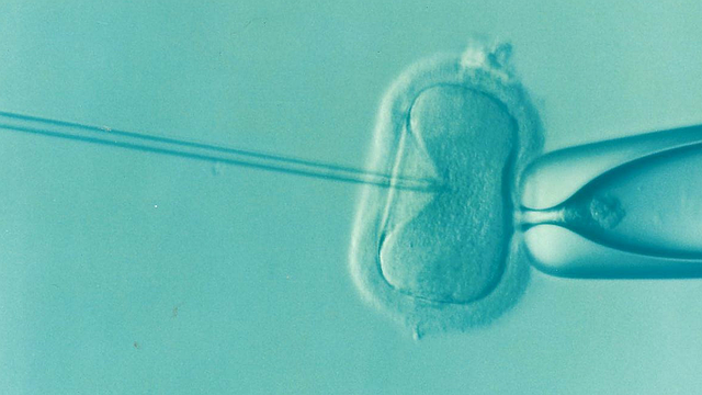 A common clinical scenario: a couple with male factor infertility (varicocele with poor semen parameters) is referred directly to an IVF clinic without evaluation for surgical repair. The rationale is efficiency — “bypass the problem with IVF.” The data, however, suggest this approach is suboptimal for a substantial proportion of couples.
Varicocele repair improves IVF outcomes. A meta-analysis published in Human Reproduction found that men with varicocele who underwent surgical repair before IVF-ICSI had significantly higher fertilization rates, embryo quality, and clinical pregnancy rates compared to men with untreated varicocele proceeding to IVF-ICSI. The DNA fragmentation improvement after varicocelectomy is the likely driver — ICSI uses individual sperm and cannot compensate for the downstream consequences of high DFI on embryo development.
Additionally, varicocele repair before IVF allows some couples to achieve natural conception, avoiding the cost, burden, and risk of IVF entirely. In a couple where the female partner is under 35 with good ovarian reserve, the expected natural conception rate of 36-45% within 24 months after varicocelectomy may compare favorably with IVF both economically and biologically.
For couples with time pressure (female partner over 38, declining ovarian reserve) or where semen parameters are severely compromised (azoospermia requiring TESE), proceeding with IVF without waiting for surgical recovery is often the right choice. But the default approach of skipping surgical evaluation entirely in favor of immediate IVF misses an opportunity to improve outcomes and in many cases to achieve a natural result.
POST-OPERATIVE CARE AND RECOVERY
Microsurgical subinguinal varicocelectomy is typically performed as an outpatient procedure under general or spinal anesthesia, with total operative time of 60-120 minutes for unilateral and 90-180 minutes for bilateral repair. The incision is small (2-3 cm) at the subinguinal level.
Recovery protocol varies by surgeon, but generally: scrotal support with athletic supporter for 4-6 weeks. Avoid heavy lifting (more than 10 lbs) and strenuous activity for 3-4 weeks. Most men return to office work within 1-2 days and physical labor within 2-3 weeks. Sexual activity can typically resume at 7-10 days.
Complications of microsurgical varicocelectomy are uncommon. Hydrocele (fluid accumulation around the testis, from inadvertent ligation of lymphatics) occurs in approximately 1% of cases with the microsurgical approach (versus 5-10% with non-microsurgical techniques). Testicular artery injury with resultant testicular atrophy is exceedingly rare (<0.5%) with microsurgical technique. Wound infection and hematoma are infrequent with standard surgical precautions.
Post-operative semen analysis should be performed at three months and again at six to twelve months. Hormonal testing at six months is appropriate for men who had pre-operative testosterone deficiency. If semen parameters have not adequately improved at twelve months and fertility remains the goal, reassessment with ultrasound to evaluate for recurrence and consideration of assisted reproductive technology is the appropriate next step.
Anatomy Varicocele WhatS: Your Questions Answered
Q: Can a varicocele be treated without surgery?
A: Non-surgical medical management — including antioxidant supplementation and lifestyle modification — can partially mitigate the oxidative damage caused by varicocele but cannot correct the underlying anatomical problem. A varicocele is a structural abnormality (venous valve failure) that does not self-correct. Antioxidant therapy (particularly CoQ10, vitamin C, vitamin E, and NAC) has been shown to improve semen parameters and reduce DNA fragmentation in men with varicocele, sometimes meaningfully, but it does not address the thermal and hemodynamic mechanisms of damage.
For men with mild effects and normal semen parameters, conservative management is appropriate. For men with significant fertility impact, pain, or hypogonadism, structural correction (surgical or radiological) is the evidence-based approach.
Q: Will my semen analysis return to completely normal after varicocelectomy?
A: Not necessarily, and this depends heavily on the baseline parameters and the duration and grade of the varicocele. Men with moderate impairment (count 5-20 million/mL, motility 20-40%) frequently normalize or achieve clinically adequate parameters after repair. Men with severe impairment (count below 5 million/mL, particularly with elevated FSH) may see meaningful improvement without reaching the normal reference range.
The clinical goal is not achieving a perfect semen analysis but achieving parameters compatible with natural conception or improving parameters enough to enhance IVF success rates. Even partial improvement matters enormously for outcomes.
Q: Does varicocele affect one testis or both?
A: While 90% of varicoceles are left-sided clinically, both testes are frequently affected functionally. The left testicular vein drainage impairment causes venous hypertension that propagates through the connecting venous plexuses to affect right testicular temperature and blood flow. This “bilateral effect” from a unilateral varicocele explains why bilateral semen parameter improvements are seen after left-sided repair alone. Truly bilateral clinical varicoceles (palpable on both sides) are found in about 10% of cases and are treated by repairing both sides.
Q: Is there a risk that varicocele repair will cause permanent damage to the testis?
A: The risk of significant testicular damage from microsurgical varicocelectomy is extremely low (<0.5%) when performed by an experienced microsurgeon. Testicular artery injury is the most feared complication, but under magnification the arterial structures are clearly identifiable and preserved. Choosing a urologist or andrologist with dedicated microsurgical training and high case volume substantially reduces this risk.
The risk of NOT treating a symptomatic varicocele — progressive testicular atrophy, worsening testosterone, and accumulating fertility damage — typically outweighs the surgical risk in appropriate candidates.
Q: My doctor says my varicocele is “subclinical” on ultrasound only. Should I still treat it?
A: Current evidence does not support treating subclinical varicoceles for fertility purposes. The AUA and ASRM guidelines specifically recommend against it. Subclinical varicoceles do not predictably improve semen parameters with treatment, and the published RCT data shows no benefit over observation in this group. The indication for treatment requires a palpable varicocele on physical examination, documented infertility, and adequate female evaluation.
If unexplained infertility exists alongside an ultrasound-only finding, the investigation should focus on other potential contributors rather than treating the subclinical varicocele.
Q: How long after varicocelectomy should we wait before trying to conceive?
A: There is no biological reason to delay attempting conception after varicocelectomy — semen parameters start improving immediately as the thermal and oxidative stressors are removed, and natural attempts can continue throughout the recovery period. However, the practical advice is to allow 3-6 months before drawing conclusions about whether the surgery has been effective. The full semen parameter benefit typically takes 6-12 months to manifest.
If the female partner’s age and ovarian reserve permit, a six-month post-operative observation period before moving to assisted reproduction is reasonable for most couples.
THE BIGGER PICTURE: VARICOCELE AS A SYSTEMIC HEALTH INDICATOR
Varicocele is not merely a fertility problem — it is a marker of systemic vascular vulnerability. Men who develop varicocele in their twenties are exhibiting venous valve incompetence at a relatively young age. The same underlying tendency toward venous insufficiency predicts higher lifetime risk of varicose veins in the lower extremities, hemorrhoids, and potentially other venous competence disorders.
The testosterone deficiency that often accompanies untreated varicocele carries its own long-term health implications. Hypogonadism is associated with increased cardiovascular risk, reduced bone mineral density, impaired glucose metabolism, and accelerated cognitive aging. A varicocele that is dismissed as a minor anatomical variant may be quietly contributing to a hormonal environment that accelerates multiple aging processes across decades.
This framing matters because it changes the calculus of treatment. For a 24-year-old man with a grade III left varicocele, the question is not merely “will repairing this help me have children faster” — it is “what does leaving this untreated do to testosterone, testicular health, and overall metabolic trajectory over the next forty years?” The data suggest the answer is: nothing good.
Derek had his varicocele repaired by microsurgery at thirty years old. At the three-month semen analysis, his total motile count had more than doubled. At twelve months, his testosterone, which had been borderline low, was firmly in the normal range. His wife conceived naturally eight months after his surgery. He occasionally thinks about those three years he spent telling himself the ache was nothing.
The most expensive medical care is the care delayed because nothing seemed wrong.
VARICOCELE AND THE FERTILITY TIMELINE: MAKING THE DECISION
For couples navigating infertility with a male varicocele diagnosis, the central clinical question becomes: repair and wait, or proceed to assisted reproduction now? The answer is never universal and always depends on specific clinical variables. A framework for working through this decision:
First, assess the female partner’s fertility status comprehensively. Ovarian reserve (AMH, antral follicle count), tubal patency, uterine anatomy, and age are the critical variables. A female partner with excellent ovarian reserve (AMH above 2.0 ng/mL) and normal anatomy at age 30 can afford to wait 12 months for varicocelectomy recovery without meaningful fertility loss.
A female partner with AMH of 0.8 ng/mL at age 38 has a very different calculus — time is genuinely the enemy, and the decision tilts toward concurrent IVF while repairing the varicocele for long-term testicular health.
Second, assess the severity of the male factor. Grade III varicocele with total motile count of 8 million and DFI of 32% in a man with normal FSH is an excellent surgical candidate with a strong predicted response. The same varicocele grade in a man with FSH of 30 mIU/mL, count of 0.5 million, and testicular volume of 8 mL bilateral is a much poorer surgical candidate — the testicular damage may be too advanced for meaningful recovery.
Third, consider the couple’s preferences and resources. IVF is expensive, physically demanding for the female partner, and carries small but real medical risks (ovarian hyperstimulation syndrome, multiple pregnancy, procedural risks). For couples who prefer to maximize natural conception probability and have the time to pursue surgical recovery, varicocelectomy followed by timed natural attempts is often the more satisfying path when clinically appropriate.
For couples who have been trying for three or more years and have significant emotional exhaustion, the faster timeline of IVF may matter more than the theoretical benefits of surgery first.
A practical synthesis: for couples where the female partner is under 36 with good ovarian reserve, the male factor is varicocele with moderate impairment, and the varicocele is high-grade and palpable, varicocelectomy followed by 9-12 months of natural attempts is the first-line recommendation of most reproductive urologists and is consistent with the available evidence on outcomes. For all other scenarios, individualized decision-making with both a reproductive urologist and a reproductive endocrinologist is essential.
The field of male fertility management is slowly shifting from a default of “treat the woman, assist the conception” toward recognizing male reproductive health as a treatable system with its own pathophysiology and intervention opportunities. Varicocele sits at the center of that shift — a condition that is common, identifiable, treatable, and consequential, that has been chronically undertreated because the men who have it were never systematically told to care about it.
That knowledge gap is the actual problem. The biology has been understood for decades. The interventions work. What fails is the system of information, examination, and advocacy that should be routing men toward evaluation in the first place.
The genetic and epigenetic consequences of high sperm DNA fragmentation extend beyond the couple’s fertility journey. Emerging research suggests that children conceived through IVF with sperm having high DFI may have subtle differences in DNA methylation patterns that affect developmental regulation.
While the long-term clinical significance of these epigenetic differences remains under investigation, the principle is clear: optimizing paternal sperm DNA integrity before conception is not merely about getting pregnant — it is about the quality of the genetic material that seeds a new human life. This reframes male fertility optimization from a narrow clinical problem into something closer to a fundamental act of parental responsibility. The testis is not a passive organ that either works or doesn’t.
It is a dynamic system that responds to inputs — thermal, oxidative, hormonal, nutritional — over the 74-day spermatogenic cycle. Every cycle is an opportunity to improve the output. Varicocele removes that opportunity systematically, heat-by-heat and radical-by-radical, until the cumulative damage becomes unmistakable in a semen analysis or a fertility clinic’s statistics. Treatment restores the opportunity.
That is what the surgery ultimately does: it removes the hand that has been pressing down on the scale, and lets the system find its own level again.
The Mechanisms That Drive Anatomy Varicocele WhatS
Understanding the biological mechanisms underlying varicocele transforms the approach from guesswork to precision. The surface-level advice — do this, avoid that — is useful as a starting point but insufficient for optimization. The men who achieve the best outcomes are the ones who understand why a protocol works, which allows them to troubleshoot when it doesn’t and adapt when their circumstances change.
At the cellular level, the processes involved are governed by signaling cascades that respond to environmental inputs — diet, movement, sleep, and stressors encountered. These cascades are not static; they adapt over days to weeks based on the signals they receive. This is why a protocol that works for the first month may lose effectiveness: the biology has adapted to the stimulus, and the signal needs to change. Periodization — the systematic variation of stimulus over time — is not just a training concept. It applies to nutrition, supplementation, stress management, and virtually every other health intervention.
The inflammatory dimension deserves particular attention. Chronic low-grade inflammation — sometimes called inflammaging when it occurs in the context of biological aging — is implicated in virtually every chronic disease state relevant here. The markers most clinicians track (CRP, ESR) capture only the most obvious systemic inflammation. More sensitive markers — including IL-6, TNF-alpha, and oxidized LDL — often reveal inflammatory activity that standard testing misses entirely. Normal-looking standard labs alongside symptoms that don’t feel normal is frequently where inflammatory markers reveal the discrepancy.
How Varicocele Disrupts Your Hormones
Hormones are not isolated actors — they operate in cascades where upstream changes propagate downstream through multiple systems simultaneously. The hormonal context matters enormously here. Cortisol dysregulation alone can explain symptoms ranging from fatigue and weight gain to poor sleep and cognitive decline — all of which may be attributed to other causes if cortisol is never measured.
The cortisol-testosterone relationship is particularly relevant for men. Chronic cortisol elevation suppresses testosterone production through the pregnenolone steal mechanism — the shared precursor is diverted toward cortisol at the expense of testosterone, DHEA, and progesterone. A man with low testosterone may not have a testicular problem at all — the underlying issue may be a stress problem manifesting hormonally. Treating the testosterone without addressing the cortisol treats the effect while ignoring the cause.
Thyroid function adds another layer. The conversion of T4 to active T3 occurs primarily in the liver and gut — not in the thyroid itself. This means that liver health, gut health, and nutrient status (particularly selenium, zinc, and iron) all influence effective thyroid function. A standard TSH test may read as normal while function is effectively hypothyroid because the conversion process is impaired. Comprehensive thyroid panels that include free T3, free T4, reverse T3, and TPO antibodies — not just TSH — give a fuller picture. See the diagnostics hub for the complete testing framework.
Your Varicocele Action Plan
A protocol for addressing varicocele-related fertility impairment should be built in phases, not implemented all at once. Phase one — typically weeks one through four — establishes the foundation: sleep optimization, dietary cleanup (removing processed foods and inflammatory seed oils), basic supplementation (vitamin D, magnesium, omega-3), and daily movement. Phase two — weeks five through eight — adds targeted interventions based on specific lab work and symptom profile. Phase three — weeks nine through twelve and beyond — introduces advanced protocols and fine-tuning based on response data.
The most common mistake is attempting Phase three interventions without completing Phase one. Advanced protocols — whether they involve peptides, specialized supplementation, or intensive training programs — assume a functioning biological foundation. Without adequate sleep, basic nutrition, and stress management, these interventions either fail to produce expected results or produce paradoxical effects that create confusion and frustration.
For personalized guidance on where to start, the interactive assessment tools help identify a specific baseline. For the complete evidence base, explore the topic directory. And for the conversational depth that written articles cannot fully capture, the podcast archive covers many of these topics across 395 episodes.
Testicular Oxidative Stress: The Central Mechanism of Varicocele-Related Damage
Understanding why varicoceles damage spermatogenesis requires moving from the macroscopic anatomy of dilated veins down to the cellular and molecular level where the actual damage occurs. The central mechanism — now supported by decades of research and confirmed in multiple independent laboratory models — is oxidative stress: an imbalance between reactive oxygen species (ROS) production and antioxidant defense capacity in the testicular microenvironment that directly impairs sperm development and DNA integrity.
Reactive oxygen species are produced as normal byproducts of cellular metabolism. In controlled amounts, they serve as signaling molecules. In excess, they damage lipids, proteins, and nucleic acids. The testis is particularly vulnerable to oxidative damage for two reasons: sperm cells have extremely high concentrations of polyunsaturated fatty acids in their membranes — these long-chain fats are essential for membrane fluidity and the acrosome reaction but are highly susceptible to lipid peroxidation — and developing spermatocytes have reduced antioxidant enzyme activity compared to most somatic cells.
The testicular environment is therefore finely balanced between the ROS required for normal sperm function and the antioxidant capacity needed to prevent oxidative damage.
Varicoceles disrupt this balance in multiple ways. Venous stasis allows hypoxic blood to pool in testicular veins, and hypoxic conditions stimulate ROS production through incomplete mitochondrial electron transport. The elevated testicular temperature from impaired countercurrent heat exchange increases metabolic rate and ROS generation. Reflux of renal and adrenal metabolites — particularly catecholamines, cortisol metabolites, and prostaglandins from the left renal vein into the left testicular vein — introduces molecules that independently generate oxidative stress in testicular tissue. The result is a microenvironment with chronically elevated ROS that overwhelms local antioxidant defenses.
The cellular consequences are multiple and cumulative. Spermatogonial stem cells — the earliest cells in the spermatogenic lineage — show reduced proliferation rates in varicocele models, indicating that the damage begins before the commitment to meiosis. Primary spermatocytes show increased rates of meiotic arrest. Spermatids — the post-meiotic cells that elongate and develop the flagellum and acrosome — show oxidative damage to their mitochondrial DNA, which provides the ATP for sperm motility. Mature spermatozoa collected from men with varicoceles show elevated 8-hydroxydeoxyguanosine in nuclear DNA, which is a direct biomarker of oxidative DNA damage. This sperm DNA fragmentation — measurable with the DNA fragmentation index (DFI) — impairs fertilization potential and embryo development independently of conventional sperm parameters.
The antioxidant supplementation research in varicocele has been partly motivated by this oxidative mechanism. Coenzyme Q10, vitamin C, vitamin E, NAC (N-acetyl cysteine), and astaxanthin have all been studied individually and in combination in men with varicoceles, with generally positive effects on sperm parameters. The combination of antioxidant supplementation with varicocele repair (microsurgical varicocelectomy) appears to produce better outcomes than either intervention alone — consistent with the hypothesis that the surgery removes the source of excess ROS while antioxidants support recovery of the spermatogenic epithelium that has already been damaged.
The Bilateral Question: Why Unilateral Varicocele Affects Both Testes
One of the most diagnostically and therapeutically important observations in varicocele biology is that the testicular damage is frequently bilateral, even when only one varicocele is clinically detected. A man diagnosed with a left-sided varicocele — which is the case in the vast majority of varicocele presentations — often shows impaired spermatogenesis and reduced testicular function in both testes, not just the left. Understanding this bilateral effect requires understanding the cross-talk mechanisms between the two testes and the systemic effects of varicocele pathophysiology.
Three mechanisms explain the contralateral effect. First, the venous anatomy of the scrotum includes communicating veins between the left and right testicular venous systems within the pampiniform plexus. Elevated venous pressure on the left can be transmitted to the right through these communicating branches, producing subclinical venous stasis in the right testis even without a clinically visible right varicocele. Second, the elevated circulating reactive oxygen species and metabolite reflux from a left varicocele enter the systemic circulation and can reach the right testis through the arterial supply. The right testis is downstream of the same systemic oxidative stress produced by the left varicocele’s venous pooling. Third, the systemic endocrine effects of varicocele-related testicular dysfunction — particularly the alterations in testosterone production and LH/FSH regulation — affect both testes simultaneously through shared hypothalamic-pituitary regulation.
This bilateral pathophysiology explains several clinical observations. Men with clinically unilateral left varicoceles often show symmetric testicular volume loss — both testes are smaller than expected for age, not just the left. Semen analysis abnormalities (reduced motility, elevated morphology defects, increased DNA fragmentation) reflect the combined output of both testes and are therefore worse than would be expected if only the left testis were affected. After varicocele repair, semen parameter improvements are often contributed to by improved right testicular function — a contralateral benefit that can only be explained if the right testis was suppressed by the left-sided varicocele even without direct structural abnormality.
The bilateral question also has clinical implications for the controversial right-sided varicocele. Right varicoceles are rare (five to ten percent of clinical varicoceles) because the right testicular vein drains directly into the inferior vena cava at a shallow angle that makes reflux less likely than on the left. When a right varicocele is found, particularly in isolation, it prompts investigation for retroperitoneal pathology — a right-sided renal mass or lymph node enlargement compressing the right testicular vein is a recognized cause of right varicocele formation that requires exclusion before attributing the finding to primary venous incompetence. This clinical caution — right varicocele warrants imaging — is a practical diagnostic point with patient safety implications.
Varicocele and Testosterone: The Endocrine Dimension
The relationship between varicocele and testosterone levels has generated considerable research interest and some genuine controversy. Varicoceles are associated with reduced Leydig cell function — the testosterone-producing cells of the testis — in a substantial proportion of affected men, and varicocele repair has been shown in several studies to improve testosterone levels in hypogonadal men with confirmed varicoceles. This positions varicocele not merely as a fertility issue but as a potentially treatable cause of hypogonadism in men who might otherwise receive testosterone replacement therapy as a default management strategy.
The Leydig cell damage mechanism parallels the Sertoli cell and spermatogenic damage: elevated temperature, oxidative stress, and hypoxia in the testicular microenvironment impair Leydig cell steroidogenesis. Leydig cells require intact mitochondrial function for the rate-limiting step of cholesterol transport into the inner mitochondrial membrane — the step mediated by the StAR protein that initiates testosterone synthesis. Mitochondrial oxidative damage from varicocele-associated ROS directly impairs this process. Additionally, elevated intratesticular temperature reduces the expression of the LH receptor on Leydig cells, making them less responsive to pituitary LH stimulation even when hypothalamic-pituitary function is intact.
The clinical data on varicocele repair and testosterone are particularly relevant for men presenting with hypogonadism symptoms and a confirmed varicocele. A systematic review by Tanrikut and colleagues found mean testosterone increases of approximately 100-150 ng/dL following microsurgical varicocelectomy in hypogonadal men with varicoceles — modest but clinically meaningful improvements that brought some men from frankly hypogonadal levels into the low-normal range. For men with borderline testosterone who are considering whether to initiate testosterone replacement therapy (which suppresses endogenous testosterone production and typically renders men infertile while on therapy), varicocele repair represents a potentially superior first intervention: it addresses the underlying cause rather than compensating for the consequence, and it preserves fertility potential that exogenous testosterone would compromise.
The intersection of varicocele management and testosterone replacement therapy planning is an increasingly important clinical conversation as both conditions become more commonly recognized and addressed. A man in his thirties or forties presenting with fatigue, reduced libido, and testosterone levels in the 250-350 ng/dL range — below the population mean but within the normal reference range — deserves a scrotal ultrasound as part of his workup if varicocele hasn’t been excluded. Finding and treating a grade II or III varicocele in this clinical context may achieve the therapeutic goal of improved testosterone function without committing the patient to lifelong exogenous hormone dependence and the attendant fertility suppression.
References
Editorial StandardsCorrectionsMedical DisclaimerAbout Our ContentAffiliate DisclosureSite Map