
Shoulder impingement is the most commonly diagnosed shoulder condition in orthopedic practice, and the surgery used to treat it performs no better than sham operations in controlled trials. The research on this point is, at this stage, overwhelming. What actually works is a targeted rehabilitation protocol that most patients never receive — because it requires understanding the biomechanics that caused the impingement in the first place, and that’s not a five-minute conversation in an orthopedic clinic.
What nobody told Derek at that appointment was that the research on shoulder impingement surgery versus conservative rehabilitation is, at this point, brutally one-sided. A 2015 systematic review by Jeremy Lewis — one of the most-cited physiotherapists in the shoulder literature — concluded that structured exercise rehabilitation for subacromial impingement produces outcomes equivalent to surgery, with significantly less risk, no recovery time, and a fraction of the cost. Exercise, as effective as surgery. For a condition affecting millions of people. Largely unknown outside specialist circles.
Derek did the rehabilitation program. Eight weeks. His pain went from a 7/10 to a 1/10. He returned to full overhead work. No surgery.

What Shoulder Impingement Actually Is
Subacromial shoulder impingement is the most common cause of shoulder pain, accounting for roughly 44-65% of all shoulder pain presentations in clinical settings. The term itself is somewhat contested in modern sports medicine — many clinicians prefer “rotator cuff tendinopathy” or “subacromial pain syndrome” — but the underlying mechanics are consistent enough to discuss under one unified model.
The basic anatomy: the shoulder joint is a ball-and-socket joint, the head of the humerus (upper arm bone) articulating with the glenoid fossa of the scapula. Above the joint sits a bony roof, the acromion. Between the humeral head and the acromion runs the subacromial space — a narrow corridor housing the supraspinatus tendon (part of the rotator cuff), the subacromial bursa, and the long head of the biceps tendon.
Impingement happens when that space narrows dynamically during shoulder movement, compressing the supraspinatus tendon and bursa against the underside of the acromion. The compression creates inflammation, pain, and, over time, tendon degeneration and potential partial or full tears.
The key word there is dynamically. The subacromial space narrows during shoulder elevation — particularly between 60 and 120 degrees of abduction, the “painful arc.” In a normally functioning shoulder, the rotator cuff muscles (supraspinatus, infraspinatus, teres minor, subscapularis) work together to depress the humeral head, keeping subacromial clearance intact even as the arm rises. When the rotator cuff is weak, fatigued, or poorly coordinated, the humeral head fails to depress adequately and rides upward instead — compressing everything above it. That’s the mechanical failure that produces impingement.
There’s a second mechanism too. Scapular dyskinesis — abnormal scapular positioning and movement — reduces the available space differently: instead of the humeral head rising too high, the acromial roof drops too low. Poor scapular upward rotation during arm elevation, caused by weak serratus anterior and lower trapezius, cuts the clearance from above. Most shoulder impingement cases involve both mechanisms to some degree.
The Diagnostic Assessment: What You Can Check Yourself
Definitive diagnosis requires a physiotherapist or sports medicine physician. But several self-assessment tools give a strong indication of whether impingement is the issue, versus other shoulder pathology — frozen shoulder, AC joint problems, labral tears, cervicogenic referred pain.
The Painful Arc Test: Stand with the arm at the side. Slowly raise it sideways (abduction) from 0 to 180 degrees. Pain occurring specifically between 60 and 120 degrees, and diminishing above 120, is highly indicative of subacromial impingement. Pain throughout the whole arc suggests something else. No pain at all — good sign.
The Empty Can Test: Raise the arm to 90 degrees in the scapular plane (about 30-40 degrees forward of straight sideways). Thumb turned down, as if emptying a can. Have someone push down on the arm while resisting. Pain or weakness here is a positive test, indicating supraspinatus involvement. Significant weakness — the arm gives way — suggests a possible rotator cuff tear rather than simple impingement.
The Hawkins-Kennedy Test: Raise the arm forward to 90 degrees, then have someone internally rotate it — turning the hand toward the floor. Pain with this maneuver is a positive Hawkins-Kennedy sign, indicating subacromial impingement. This test has a sensitivity of approximately 79% — it catches most true impingement cases, though there are some false positives.
The Drop Arm Test: Screens for significant rotator cuff tears. Passively raise the arm to 90 degrees abduction, then lower it slowly. Inability to control the lowering, or the arm dropping suddenly, suggests a significant rotator cuff tear that may need surgical consultation regardless of other findings.
Positive Painful Arc and Hawkins-Kennedy results without significant weakness — good candidate for the conservative rehabilitation approach below. Significant weakness, or a positive Drop Arm test — get imaging and a specialist opinion before proceeding with anything else.
The Lewis 2015 Research: Why Surgery Is Usually Not the Answer
Jeremy Lewis’s 2015 systematic review in the journal British Journal of Sports Medicine examined the evidence for subacromial decompression surgery — acromioplasty, a procedure that shaves the underside of the acromion to create more space — versus conservative treatment for subacromial impingement.
The findings were unambiguous. Exercise-based rehabilitation produced outcomes equivalent to surgery at every follow-up time point measured: 6 weeks, 3 months, 6 months, 12 months. No statistically significant difference in pain, function, or quality of life between surgical and conservative groups. A landmark 2018 Finnish randomized controlled trial (the FIMPACT trial, published in BMJ) went further still, finding that sham surgery — an incision under anesthesia with no actual decompression performed — produced the same outcomes as real acromioplasty. The surgery was no better than a placebo procedure. That’s worth sitting with for a second.
None of this means shoulder surgery is never warranted. Large full-thickness rotator cuff tears that don’t respond to rehabilitation, significant instability from labral tears, certain structural abnormalities — these are legitimate surgical indications. But for the common presentation of subacromial impingement in an otherwise healthy adult, the evidence strongly favors exhausting conservative rehabilitation before any surgical intervention gets considered.
“There is no evidence that acromioplasty is more effective than exercise rehabilitation for subacromial shoulder pain. For most patients, the surgical option should be reserved for those who have undergone a genuine trial of rehabilitation and failed to achieve acceptable outcomes.” — Lewis JS, British Journal of Sports Medicine, 2015
Phase 1: Pain Management and Motor Control (Weeks 1-2)
The Shoulder Rehabilitation Ladder is a phased approach that progressively loads the rotator cuff and periscapular muscles, starting from pain-free isometric contractions and building up to full overhead loading. Each phase has to be completed before moving to the next one. Skipping phases is the single most common reason shoulder rehabilitation programs fail.
Load management: In the acute phase, avoid anything that reproduces pain above a 3/10. That means temporarily modifying overhead work, not sleeping on the affected shoulder, stopping any upper body exercise that causes pain. The goal isn’t immobilizing the shoulder — movement is what heals it — but reducing the inflammatory load enough that the exercises can actually be performed without provoking a flare-up.
Pendulum exercises: Lean forward, support with the unaffected arm on a table, let the affected arm hang. Gently swing it in small circles, forward and back, side to side — using gravity and momentum, not active muscle contraction. This creates gentle joint distraction and promotes synovial fluid circulation in the joint. 2 minutes, 3x daily.
Scapular setting: Sit or stand upright. Gently draw the shoulder blades down and slightly back — not a military “attention” posture, just a gentle depression and retraction. Hold 5 seconds, release. 15 repetitions, 2x daily. This activates the lower trapezius and starts re-educating scapular positioning.
Isometric rotator cuff activation: Stand next to a wall, elbow bent at 90 degrees and pressed against the wall. Gently push outward (external rotation) without moving. Hold 5 seconds, 10 repetitions. Then push inward (internal rotation). 10 repetitions each direction, 2x daily. These pain-free isometrics start restoring the force-couple relationship of the rotator cuff without creating the dynamic compression that the impingement arc produces.
Phase 2: Rotator Cuff Strengthening (Weeks 3-5)
Once all Phase 1 exercises can be done pain-free and resting shoulder pain sits below 2/10, move to Phase 2. This phase focuses on the specific rotator cuff strengthening that creates the humeral head depression needed to maintain subacromial clearance.
Side-lying external rotation: Lie on the unaffected side, affected arm resting on the body, elbow bent at 90 degrees. With a light resistance band or a 1-2kg dumbbell, externally rotate the forearm upward, elbow tucked to the side. Lower slowly. 3 sets of 15. This specifically trains the infraspinatus and teres minor — the primary external rotators and key humeral head depressors. Most shoulder impingement cases involve weakness specifically in external rotation.
Prone Y-T-W: Lie face down, arms extended overhead (Y position), out to the sides (T position), or bent at 90 degrees (W position). Lift each position a few centimetres off the ground, hold 3 seconds. No weight needed at first — bodyweight is challenging enough in these positions. 3 sets of 10 each. These target the lower trapezius and serratus anterior specifically, the muscles responsible for scapular upward rotation during arm elevation.
Banded pull-apart: Hold a resistance band with both hands in front, arms extended, shoulder-width grip. Pull the band apart horizontally until the hands reach the sides. Return slowly. 3 sets of 15. Trains the posterior deltoid, rhomboids, and mid trapezius — all important for scapular stability and shoulder retraction.
Wall slide: Face a wall, forearms pressed against it, elbows at shoulder height. Slowly slide the arms upward, staying in contact with the wall, until fully extended overhead. Return. Trains scapular upward rotation through a controlled range. 3 sets of 10. Progress by extending wall contact to include the upper arms as mobility improves.
Phase 3: Thoracic Mobility — The Missing Link
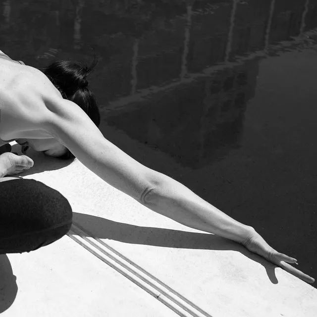
Thoracic extension over foam roller: Place a foam roller perpendicular to the spine at the mid-back. Support the head with the hands. Gently extend over the roller, letting gravity mobilize the thoracic segments. Move the roller up and down the mid-back (T3-T9 region). 60-90 seconds daily. The most efficient thoracic mobility tool in the rehabilitation toolkit, arguably.
Open book rotation: Lie on the side, knees bent at 90 degrees. Extend the top arm forward, stacked on the bottom arm. Slowly rotate the top arm backward, opening the chest toward the ceiling, eyes following the hand. Hold at end range 2-3 seconds. Return. 10 reps per side. This restores thoracic rotation, which is independently useful and also facilitates better scapular motion during arm elevation.
Chin tuck with thoracic extension: Standing against a wall, perform a chin tuck (draw the head straight back, not up or down) while simultaneously pressing the thoracic spine gently against the wall. Hold 5 seconds, 10 repetitions. This corrects forward head posture and thoracic kyphosis at the same time, which directly reduces the mechanical stress on the subacromial space by changing the acromion’s relative position.
Phase 4: Progressive Loading (Weeks 6-8)
Once rotator cuff strength has been established and thoracic mobility has improved, the shoulder needs progressive loading to build the strength and tissue tolerance necessary to return to full activity — particularly overhead activity like Derek’s electrical work.
The progression follows a load-tolerance model: tissue heals and adapts in proportion to the load applied to it, as long as the load stays below the threshold that triggers inflammation. Phase 4’s whole job is systematically raising that threshold.
Dumbbell lateral raise (pain-free range): Start in the pain-free range (typically 0-60 degrees) with a light weight. Progressively increase both weight and range over the weeks. By the end of Phase 4, most people can complete a full lateral raise to shoulder height without pain. Key rule: any rep producing pain above a 3/10 stops the set. Pain during exercise up to 3/10, and persisting up to 24 hours post-session, is acceptable — normal tissue loading. Pain above 3/10, or lasting more than 24 hours, means the tissue’s current tolerance has been exceeded.
Cable face pull: Using a cable machine or resistance band at eye level, pull the handles toward the face, driving the elbows out and up. Loads the posterior rotator cuff and rear deltoid in a scapular plane position that minimizes impingement while building the strength needed for overhead activity. 3 sets of 15.
Landmine press (shoulder-safe overhead press progression): Instead of a standard overhead press, which places heavy demand on scapular upward rotation and can provoke symptoms, the landmine press — pressing at a 45-degree angle with a barbell anchored in a corner — provides overhead loading in a position that’s mechanically forgiving to the shoulder. Progress from here toward a full overhead press as symptoms allow.
Long-Term Prevention: Building a Shoulder That Doesn’t Break
Shoulder impingement recurrence is common, because people finish rehabilitation, return to normal activity, and slowly recreate the exact conditions that produced the impingement the first time — weak external rotators, tight pectorals, poor thoracic mobility, excessive internal rotation dominance.
Prevention means addressing the root causes. Most modern adults have a systematic imbalance between anterior (internal rotation) and posterior (external rotation) shoulder strength. We push, type, drive. We rarely pull in the horizontal plane or externally rotate against resistance. In a well-designed training program the ratio of pulling to pushing should sit at minimum 2:1 — two pulling exercises for every pressing exercise. Most people who develop shoulder impingement are running ratios closer to 1:1, or even 1:2.
The maintenance protocol for long-term shoulder health is simple enough: horizontal pulling (rows) and vertical pulling (pull-downs, pull-ups) in every upper body training session. Band pull-aparts or face pulls as a warm-up. Thoracic mobility maintained with foam rolling 2-3x a week. Avoid sleeping on the affected shoulder — or any shoulder, really — for extended periods, since prolonged compression of the subacromial space during sleep is an underappreciated contributor to morning shoulder pain.
Occupational considerations matter too. Work involving repetitive overhead motion — electricians, painters, plasterers, welders — keeps the rotator cuff under constant eccentric loading that accumulates fatigue over time. Proactive strengthening of the posterior rotator cuff and periscapular muscles, not just when symptomatic but as a baseline maintenance habit, is the single most effective prevention strategy for shoulder impingement in high-risk occupations.
FAQ: Shoulder Impingement
How long does shoulder impingement take to heal with rehabilitation?
With consistent adherence to the Shoulder Rehabilitation Ladder, most people see significant pain reduction within 4-6 weeks and full or near-full function by 8-12 weeks. Cases involving significant rotator cuff tendinopathy or adhesive capsulitis may take 3-6 months. The timeline depends heavily on compliance with the phased protocol and avoiding provocative activities during the loading phases.
Is it safe to continue training with shoulder impingement?
Yes, with modifications. Avoid exercises that reproduce pain above 3/10 — typically overhead pressing, lateral raises in the painful arc, behind-the-neck movements. Substitute lower-angle alternatives (landmine press, cable work below 90 degrees) and emphasize pulling exercises, which are generally pain-free and actively therapeutic. Complete rest isn’t recommended — movement promotes healing in tendinopathy.
Is cortisone injection helpful for shoulder impingement?
Corticosteroid injections can provide short-term (4-6 week) pain relief that makes rehabilitation exercises more comfortable to perform. Not a cure, and they don’t address the mechanical causes. Research shows no long-term benefit from injection alone compared to exercise alone, but injection followed by rehabilitation may produce faster early improvement than rehabilitation on its own. If pain is severe enough to prevent Phase 1-2 exercises, an injection to facilitate rehabilitation can make sense — not as a primary treatment, just a bridge.
What is the difference between shoulder impingement and a rotator cuff tear?
Impingement is a dynamic compression syndrome without tendon disruption. A rotator cuff tear is structural damage to the tendon tissue itself. They often coexist — repeated impingement is a mechanism for rotator cuff tendon damage building up over time. Differentiating them clinically requires imaging (ultrasound or MRI). Significant weakness on the empty can test, or a positive drop arm test, suggests a tear. Full-thickness tears causing severe weakness typically need surgical repair; partial tears usually respond to rehabilitation.
Can yoga or stretching make shoulder impingement worse?
Some yoga poses can aggravate shoulder impingement — specifically poses involving end-range internal rotation (some binds and backbends) and those loading the shoulder in an impingement-prone position. Downward dog and plank positions are generally well-tolerated. The relevant question is whether a specific movement reproduces the symptoms. If it does, modify or avoid it until Phase 3-4 is complete. Thoracic mobility yoga work is actively beneficial for shoulder impingement recovery, for what it’s worth.
My impingement came back after surgery. Why?
Because acromioplasty doesn’t address the muscular causes of impingement. Even after surgical decompression, if the rotator cuff stays weak and scapular stability stays poor, the humeral head keeps migrating superiorly during arm elevation — just with slightly more clearance than before. Post-surgical shoulder impingement recurrence strongly suggests inadequate rehabilitation after the surgery. The same rotator cuff and periscapular strengthening program is the treatment, surgery or no surgery.
Can I sleep on my affected shoulder?
No — avoid sleeping on the impinged shoulder during the acute and subacute phases. Sleeping on the affected side creates sustained compression of the subacromial space, which perpetuates inflammation. Sleep on the back, or the unaffected side with the affected arm supported by a pillow. Once symptoms have reduced significantly (Phase 3-4), brief periods on the affected shoulder are usually tolerable.
The Psychological Dimension: Pain Catastrophizing and Recovery
Shoulder impingement recovery isn’t a purely mechanical process. Pain science research over the last two decades has firmly established that chronic pain — including the shoulder pain of long-standing impingement — is shaped by psychological factors in ways that can either accelerate or dramatically impede recovery. None of this makes the pain “in your head.” It just makes the pain management more complete, and more effective.
Pain catastrophizing — the tendency to ruminate on pain, magnify its significance, and feel helpless in the face of it — is one of the strongest predictors of poor outcomes in musculoskeletal rehabilitation. People who catastrophize their shoulder pain are more likely to avoid exercises causing minor discomfort, more likely to interpret normal healing pain as a sign of re-injury, and significantly more likely to remain in disability at 12-month follow-up compared to those with lower catastrophizing scores on standardized measures.
The clinical application here isn’t dismissing pain — it’s calibrating the response to it. Normal tissue loading pain, the kind felt when stretching into a new range or strengthening a deconditioned muscle, is beneficial and shouldn’t be avoided. Harmful pain — sharp, joint-located, or persisting more than 24 hours after exercise — warrants modification. Learning to tell these categories apart and respond to each appropriately, rather than treating all pain as a stop signal, dramatically improves rehabilitation outcomes. This is the pain education piece that high-quality physiotherapy programs include, and that most self-directed rehabilitation programs miss entirely.
Graded exposure — gradually and systematically reintroducing the feared movements (overhead reaching, pressing, sleeping on the affected side) at manageable intensities before full return — is both a mechanical necessity and a psychological intervention. Each successful exposure without catastrophic consequence lowers the threat value of the movement and reduces the pain amplification driven by central sensitization. The rehabilitation protocol is the vehicle. The psychology is the engine.
The Occupational Risk Factor Nobody Addresses
Shoulder impingement doesn’t just come from gym programming errors. For a large share of the population it’s an occupational injury, driven by the cumulative mechanical demands of work — and the occupational context fundamentally changes the rehabilitation strategy, because the provocative inputs keep going throughout treatment rather than stopping.
High-risk occupations: electricians and overhead laborers (sustained shoulder elevation), painters and plasterers (repetitive overhead reaching), supermarket shelf-stackers (repetitive mid-range shoulder loading), dental hygienists (sustained shoulder elevation in constrained positions), and athletes in throwing and overhead sports — baseball, tennis, swimming, volleyball. In every case, the rotator cuff gets loaded repetitively in or near the impingement arc, accumulating microtrauma faster than the tissue’s repair capacity can keep up.
The key difference in occupational impingement: load management isn’t optional. Nobody can just “avoid provocative activities” when those activities are the job. The rehabilitation strategy has to incorporate workload modification — fewer overhead repetitions per hour through technique changes, rotating tasks, structured arm rests every 20-30 minutes of sustained overhead work — combined with the standard rotator cuff and scapular stabilization program.
Ergonomic interventions matter too. For overhead workers, positioning the work object as low as the task allows reduces the elevation angle demanded. For computer workers with shoulder impingement, monitor height (at or slightly below eye level) and keyboard/mouse position (forearms roughly parallel to the floor, shoulder in neutral rotation) reduce the sustained shoulder loading that keeps symptoms going.
Athletes in overhead sports carry the added complexity of sport-specific loading patterns that need more than general rotator cuff strengthening — they need sport-specific, progressive return-to-sport protocols. A baseball pitcher’s shoulder rehabilitation looks meaningfully different from a recreational tennis player’s — not in the foundational exercises, but in the loading progression and the final stage of sport-specific conditioning. In high-level overhead athletes, the rehabilitation program should ideally be designed and monitored by a physiotherapist with specific experience in that sport.
Shoulder Anatomy Comprehensive examination: Why the Rotator Cuff Is So Vulnerable
To understand why shoulder impingement is so common and so persistent, it helps to understand the fundamental anatomical trade-off the shoulder joint represents. The shoulder is the most mobile joint in the human body — capable of 360 degrees of motion in the sagittal plane, 180 degrees of abduction, and extraordinary combined ranges that let us throw, reach, climb, swim. That mobility comes at the cost of stability. There’s no getting both for free.
The glenoid fossa — the socket half of the ball-and-socket shoulder joint — is extremely shallow. The humeral head (the ball) has a surface area roughly three times larger than the glenoid fossa it sits in. The opposite of the hip joint, where the deep acetabulum provides real bony stability. In the shoulder, bony stability is minimal. Almost all shoulder stability comes from soft tissue instead: the rotator cuff muscles, the glenohumeral ligaments, the labrum, the joint capsule.
This architecture means that when any part of the soft tissue stabilizing system fails — weakness, fatigue, injury, poor neuromuscular coordination — the humeral head can translate inappropriately in any direction. Superior migration, the mechanism behind subacromial impingement, is the most common direction of failure, because the deltoid, the large superficial shoulder muscle, creates a strong superiorly-directed force during arm elevation that the rotator cuff has to constantly counteract. When the rotator cuff comes up short — too weak, too fatigued, poorly timed — the deltoid wins, and the humerus rises into the subacromial structures above it.
The vascularity of the supraspinatus tendon adds another layer of vulnerability on top of all this. The “critical zone” of the supraspinatus tendon — roughly 1cm from its insertion on the greater tuberosity — has consistently poor blood supply across anatomical studies. This hypovascular region is precisely where most supraspinatus tears begin. Poor vascularity means poor healing capacity, which is why rotator cuff tendinopathy can be so stubborn, and why the tissue loading approach of rehabilitation — stimulating tendon healing through progressive load — is essential rather than optional. Anti-inflammatory medications and rest reduce pain. They don’t stimulate the tendon remodeling that produces long-term recovery.
Understanding Your Pain: Distinguishing Impingement from Other Shoulder Conditions
Shoulder pain is common, and subacromial impingement is common, but they aren’t the same thing. Treating all shoulder pain as impingement is a diagnostic error that leads straight to failed rehabilitation. Before committing to the Shoulder Rehabilitation Ladder, it’s worth understanding the differential diagnosis and identifying what distinguishes impingement from the other conditions that present similarly.
Adhesive capsulitis (frozen shoulder): Characterized by progressive loss of glenohumeral mobility in all planes — particularly external rotation and abduction — with severe pain early on and stiffness later. Unlike impingement, which produces pain in a specific arc, frozen shoulder restricts motion globally. The Thomas capsular pattern (most restricted external rotation, then abduction, then internal rotation) is pathognomonic. Rehabilitation differs significantly here — frozen shoulder needs aggressive mobilization and range of motion recovery rather than the rotator cuff strengthening focus of impingement rehab.
AC joint dysfunction: The acromioclavicular joint, where the clavicle meets the acromion, is a separate joint that can develop its own pathology — arthritis, sprains, separations. AC joint pain typically sits at the very top of the shoulder (the bony bump) and gets reproduced by horizontal adduction (crossing the arm across the body) rather than the painful arc of impingement. Direct palpation of the AC joint is tender. The rehabilitation approach differs from rotator cuff-focused impingement rehab.
SLAP tear (Superior Labrum Anterior to Posterior): Labral tears at the top of the glenoid can produce pain similar to impingement, but with additional features — deep pain with overhead activities, a “clunking” or “catching” sensation, significant performance deterioration in throwing athletes. SLAP tears get diagnosed definitively by MRI arthrogram (a contrast-enhanced MRI). Many SLAP tears respond to conservative rehabilitation, but full-thickness tears in competitive overhead athletes may need surgical repair.
Cervical radiculopathy: Nerve root compression in the cervical spine (C5, C6, or C7 roots) refers pain to the shoulder and arm in a way that can be mistaken for shoulder pathology. Distinguishing features: pain radiating below the elbow, neurological symptoms (tingling, numbness, weakness) in specific dermatomal distributions, and symptoms reproduced by cervical movements — particularly neck extension and lateral flexion toward the affected side. Not shoulder impingement. Requires cervical spine management instead.
If the clinical picture is unclear, or standard impingement rehabilitation isn’t producing results after 4-6 weeks, a physiotherapy or sports medicine assessment is warranted. The rehabilitation approach depends entirely on getting the diagnosis right first.
Nutrition for Tendon Healing: The Supporting Role of Diet
The structural rehabilitation protocol addresses the mechanical causes of shoulder impingement. But tendon healing — the underlying tissue-level repair that determines long-term recovery — also depends on the nutritional environment surrounding it. Several specific nutrients have meaningful evidence behind them for supporting tendon metabolism and repair.
Collagen and vitamin C: Tendons are primarily composed of type I collagen. Collagen synthesis requires vitamin C as a cofactor for two enzymes (prolyl hydroxylase and lysyl hydroxylase) involved in collagen cross-linking. A 2019 randomized controlled trial by Shaw et al. demonstrated that 15g of gelatin (a hydrolyzed collagen source) taken with 50mg of vitamin C, consumed 60 minutes before exercise, significantly increased collagen synthesis markers and improved tendon mechanics compared to placebo. The timing — 60 minutes before loading — matters: the amino acids from the gelatin need time to reach the tendon before the mechanical stimulus of exercise triggers collagen deposition.
Protein adequacy: Tendon healing, like all tissue repair, needs adequate protein availability. The general recommendation for active adults in rehabilitation is 1.6-2.0g of protein per kilogram of body weight per day. Leucine, an essential amino acid abundant in animal proteins, is particularly important for triggering muscle and connective tissue protein synthesis.
Omega-3 fatty acids: EPA and DHA — the long-chain omega-3s from fish oil — have anti-inflammatory properties that may be relevant to the inflammatory component of rotator cuff tendinopathy. A 2017 study found fish oil supplementation reduced markers of tendon inflammation and improved tendon repair rates in animal models. Human evidence is less definitive, though directionally consistent. Practical recommendation: 2-4g of combined EPA/DHA daily during active rehabilitation, a reasonable, low-risk addition.
The Return to Sport Protocol: Getting Back to Full Activity
Returning to full sport or occupational activity after shoulder impingement rehabilitation requires a structured progression bridging the gap between clinical recovery and real-world loading demands. The mistake most people make is returning to full activity the moment the pain disappears — but pain disappearance indicates the inflammatory phase has resolved, not that the tissue is back to its pre-injury mechanical capacity. Tissue healing and pain resolution do not happen on the same clock.
The return to sport protocol for shoulder impingement follows a criteria-based approach rather than a time-based one. Criteria for return to overhead sport or labor: full pain-free range of motion (particularly the painful arc), rotator cuff strength testing (empty can and external rotation tests) at greater than 90% of the unaffected side, scapular stability (10 slow prone Y raises with a 1-2kg weight, no substitution), and sport-specific movement pattern testing — for throwing athletes, a graduated throwing program from 30% effort at short distance progressing to full effort over 4-6 weeks.
These criteria exist because tendons, ligaments, and bursae take longer to remodel than it takes for acute inflammation to subside. The mechanical properties of the repaired tissue — stiffness, tensile strength, fatigue resistance — aren’t fully restored until 8-12 weeks after the initial injury in most cases, longer still for significant tendinopathy. Returning to high-load overhead work before tissue maturation is complete is the most common cause of impingement recurrence — which is why so many people end up with “shoulder problems that keep coming back” despite finishing a short rehabilitation program.
The final principle of the Shoulder Rehabilitation Ladder: the top rung, preventing recurrence, is reached through strength maintenance, not through completing rehabilitation and moving on. The rotator cuff strengthening and scapular stabilization exercises developed during rehab should become permanent components of any training program involving the upper body. A shoulder that has impinged once has demonstrated a mechanical vulnerability that requires ongoing management. The management itself is simple: keep doing the face pulls, keep doing the rows, keep doing the external rotation work. The cost of maintenance is 10-15 minutes added to training sessions. The cost of recurrence — pain, restricted function, potential surgery — runs considerably higher.
References
Editorial StandardsCorrectionsMedical DisclaimerAbout Our ContentAffiliate DisclosureSite Map