
The diagnosis came after seventeen months of pain. Seventeen months of modifying runs, skipping track workouts, taping her knees the way her running group recommended, buying new shoes, stretching her IT band for twenty minutes every morning — seventeen months before a sports medicine physician finally told Diane what she actually had: patellofemoral pain syndrome, commonly called runner’s knee. Not because the diagnosis was obscure. Because the treatment path was so straightforward once correctly identified that the fact it took seventeen months of mistreatment stands as a monument to how badly recreational runners get served by generic advice, internet forums, and their own well-intentioned but fundamentally misdirected self-diagnosis.
Patellofemoral pain syndrome (PFPS) is the most common running injury, accounting for roughly 25% of all running-related injuries seen in clinical settings. It produces pain around or behind the kneecap, characteristically worsened by going down stairs, running downhill, prolonged sitting with knees bent (the “theatre sign”), and squatting. It affects more female athletes than male, peak incidence in the second and third decades of life — though it’s common at any age in anyone who runs, cycles, or does repetitive lower-extremity activity. Also one of the most common conditions seen in military recruits during basic training and in adolescents starting high-volume sports. The sheer prevalence makes understanding it well a worthwhile investment for any active person.
Despite its prevalence, runner’s knee gets routinely mismanaged because its root causes are almost never in the knee itself. The pain is in the knee. The problem is usually in the hip, the quad, or the biomechanical pattern connecting them. Treating the symptom location without addressing the mechanical source is why so many cases drag on for a year or more, when appropriately targeted rehabilitation typically produces significant improvement in 6-12 weeks.
The Anatomy: Why the Kneecap Hurts
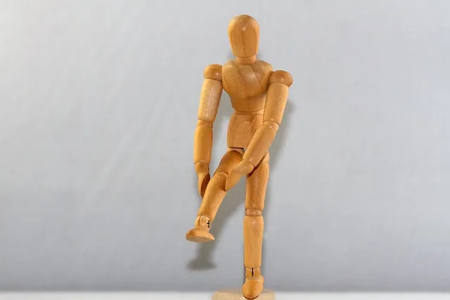
In normal mechanics, the patella tracks centrally in the groove throughout knee motion. When the forces acting on it become imbalanced — specifically when lateral forces exceed medial — the patella tracks laterally, creating increased contact pressure between the lateral facet of the patella and the lateral femoral condyle. That increased articular contact pressure, particularly under repeated loading from running, produces the characteristic anterior knee pain of PFPS.
The articular cartilage on the posterior surface of the patella doesn’t contain pain receptors — cartilage is aneural. The pain comes from the subchondral bone and the synovial tissue around the joint, which are innervated and become sensitized under abnormal, repeated mechanical stress. This is also why PFPS pain is often diffuse and poorly localized — “around the kneecap” — rather than sharply pinned to a specific structure.
The lateral tracking pattern producing PFPS is caused by mechanical contributors that can originate at multiple points along the lower extremity kinetic chain. Understanding those contributors is the key to effective rehabilitation — and explains why treating only the knee so often falls short.
The Root Causes: Why the Kneecap Misfires
Research over the past two decades has substantially revised the understanding of PFPS causation. The older model focused almost exclusively on the quadriceps — specifically the imbalance between vastus lateralis (pulling the patella laterally) and vastus medialis oblique, VMO (pulling it medially). Strengthening the VMO through terminal extension exercises was the dominant treatment approach for years.
That model is correct but incomplete. A landmark shift came from research on hip mechanics — particularly the work of Lack and colleagues (2015) and related research groups — showing hip abductor and external rotator weakness isn’t just a correlate but a likely causal contributor to PFPS. Here’s the mechanical explanation:
When the hip abductors (particularly gluteus medius) and external rotators are weak, the femur adducts and internally rotates during weight-bearing activity — running, landing, squatting. This femoral adduction and internal rotation moves the trochlear groove medially beneath a relatively fixed patella, effectively producing the same lateral tracking problem as if the patella were being pulled outward. The net effect on patellar mechanics is identical whether the patella is being pulled laterally by an overactive vastus lateralis, or the femur is moving medially beneath it.
The clinical implication is significant: rehabilitation focusing entirely on the quadriceps and ignoring the hip addresses roughly half the problem. Lack’s 2015 research comparing hip-focused rehabilitation to quad-focused rehabilitation in PFPS patients found hip strengthening produced superior outcomes — faster pain resolution, better long-term results — compared to isolated quad strengthening. This finding has been replicated enough to change clinical guidelines, though plenty of practitioners haven’t updated their practice accordingly.
Other contributing factors: IT band tightness (the lateral retinaculum attached to the IT band contributes lateral patellar tracking forces), overpronation and foot mechanics (excessive subtalar pronation rotates the tibia internally, feeding the same femoral rotation problem), patellar tendon tightness (reduces patellar mobility in the groove), and poor running mechanics — particularly excessive crossover gait, feet crossing the midline during running, which increases femoral adduction forces with each stride.
Diagnosis: What You’re Actually Dealing With
The clinical diagnosis of PFPS is primarily based on history and physical examination. Characteristic features: anterior knee pain, in front of or around the kneecap, worsened by activities involving prolonged knee flexion or high patellar contact force — squatting, running, going down stairs, prolonged sitting. The “theatre sign” — pain after sitting with knees bent more than 30 minutes that improves briefly on first standing — is particularly characteristic.
Physical examination findings typically include tenderness on patellar compression, a positive Clarke’s sign (pain with quadriceps contraction against manual patellar pressure), and often evidence of the contributing factors — hip abductor weakness demonstrable with single-leg tests (Trendelenburg sign or single-leg squat), VMO hypotrophy or timing delay on palpation, and patellar tilt (lateral patellar border higher than medial with the knee relaxed and extended).
Imaging usually isn’t necessary for diagnosis or management, but can help rule out other causes of anterior knee pain: patellar tendinopathy (pain at the inferior pole of the patella rather than diffusely behind it), fat pad impingement (Hoffa’s disease), bipartite patella, or early patellar chondromalacia. MRI is rarely necessary unless the presentation is atypical or the patient hasn’t responded to well-executed conservative management.
Distinguishing PFPS from iliotibial band syndrome (ITBS) matters clinically. ITBS produces lateral knee pain — over the lateral femoral condyle, not behind the patella — typically with a distinct onset at a specific running distance (“the 20-minute mark”), and is more associated with downhill running. PFPS pain is anterior and peripatellar. The two can coexist but call for different rehabilitation approaches.
The Knee Pain Elimination Protocol
- Clamshells: Sidelying, knees bent 90 degrees, hips flexed 45 degrees, rotate the top knee toward the ceiling while keeping feet together. 3 sets × 20 reps. Starting point for gluteus medius activation — the primary hip abductor.
- Side-lying hip abduction: Sidelying, lift the top leg with foot dorsiflexed. 3 sets × 15 reps. Progress to resisted versions with a band at the ankle.
- Hip external rotation: Seated with a resistance band around the knees, externally rotate against the band. 3 sets × 15 reps. Activates the deep hip rotators.
- Single-leg bridges: Supine, one foot flat on floor, lift hips while extending the other leg. 3 sets × 10 per side. Challenges hip abductor endurance in a weight-bearing position.
- Single-leg squats (shallow): Stand on one leg, slowly lower 20-30 degrees. Focus on a steady pelvis, avoid knee-in valgus. 3 sets × 8-10 per side. Functional hip abductor challenge under load.
This framework — the Knee Pain Elimination Protocol — is a four-phase approach organized by the evidence hierarchy for PFPS rehabilitation. It addresses the hip (primary source of biomechanical disruption), the quad (secondary contributor), the IT band and mobility (contributing structures), and the biomechanical corrections in running form that prevent recurrence.
Phase 1: Hip Strengthening (Weeks 1-6, the foundation)
Phase 2: Quad and Patellar Stabilization (Weeks 3-8, concurrent with Phase 1)
- Terminal knee extensions (TKE): Standing, resistance band behind the knee, straighten the knee from 30° flexion to full extension. 3 sets × 20 reps. Specifically activates the VMO in the range most relevant to patellar tracking.
- Step-downs: Standing on a step, slowly lower the opposite foot toward the floor while keeping the standing knee aligned over the second toe. 3 sets × 10-15 per side. Eccentric quad control under bodyweight.
- Short arc quads: Seated, rolled towel under the knee holding it at 30-45° flexion, then extend fully. 3 sets × 20 reps. VMO-targeted terminal extension.
Phase 3: Flexibility and Mobility (Ongoing from Week 1)
- Hip flexor stretching: Kneeling lunge stretch, 60-90 seconds per side. Tight hip flexors anteriorly tilt the pelvis and affect hip abductor mechanics.
- IT band/lateral hip mobility: Standing crossed-leg side-stretch, foam rolling the TFL and lateral quad, 2-3 minutes per side. Note: the IT band itself doesn’t lengthen with foam rolling, but the TFL muscle at its origin and the associated fascia benefit from release.
- Foot and ankle mobility: Ankle dorsiflexion stretching if limited — restricted dorsiflexion increases pronation during foot strike, contributing to tibial and femoral internal rotation.
Phase 4: Running Mechanics Correction (Introduction at Week 4-6)
- Cadence increase: Increasing stride rate by 5-10% (metronome or running app) reduces both stride length and vertical loading forces, decreasing patellar contact stress per stride. Most recreational runners have room to spare here.
- Midfoot strike: Transitioning from heavy heel strike to midfoot strike reduces braking force and knee flexion angle at initial contact — both reduce patellar loading.
- Hip drop prevention: Video gait analysis or a training partner watching for Trendelenburg sign (pelvis dropping on the unsupported side during running). Cue: “run tall, keep hips level.”
The knee is where runner’s knee hurts. The hip is where runner’s knee starts. Every treatment approach that ignores the hip is addressing the symptom while leaving the cause intact — and producing exactly the frustrating results that leave athletes spending months in pain wondering why the treatment isn’t working.
Running-Specific Gait Analysis: Finding Your Mechanical Contribution
Not all PFPS is driven by the same mechanical contributors. Understanding which specific factors are driving a given presentation guides more targeted rehabilitation and helps predict which parts of the protocol will produce the fastest results.
The crossover gait pattern — each foot striking near or across the midline during running — is one of the most common and treatable biomechanical contributors to PFPS. In crossover gait, lateral foot placement combined with ipsilateral hip adduction at midstance loads the lateral knee and patellofemoral joint in ways that provoke symptoms. Research shows simply cueing runners to widen their stride (step right of midline with the right foot and vice versa) significantly reduces patellofemoral loading with no other change. A quick fix that can reduce symptoms immediately while the rehabilitation program builds the strength for durable correction.
Step rate (cadence) modification is similarly accessible. Increasing cadence by 5-10% shortens stride length, reduces ground reaction force, decreases knee flexion angle at foot strike, and reduces patellofemoral contact stress per stride. A metronome app set to a target cadence, or a music playlist matched to target BPM, makes this practical during runs. Research shows this change is adoptable within 4-8 training sessions and sustainable long-term.
Hip drop (Trendelenburg) identification requires external observation or video. Film running from behind on a treadmill or track. If the pelvis drops more than 5 degrees to one side on each step — visible as an undulating “hip hike” when the supporting leg’s glute isn’t stabilizing the pelvis — that’s positive for hip abductor weakness on the supporting-leg side. This finding directly links the hip strengthening exercises to the observed movement problem, which reinforces why the hip work is the priority.
For advanced gait analysis, many running specialty clinics and sports physiotherapy practices offer 2D or 3D video analysis with slow-motion playback and joint angle quantification. A one-hour biomechanical running assessment typically reveals multiple addressable issues and provides more direction than months of generalized advice. Cost usually runs $100-250 — which competes favorably against seventeen months of pain and misdirected treatment.
Load Management During Rehabilitation
One of the most common errors in PFPS rehabilitation is complete activity cessation. Rest reduces pain temporarily because it removes the provocative loading. But tendons and cartilage need mechanical loading to maintain structural health and stimulate repair. Complete rest produces deconditioning without healing — and when running resumes, the underlying biomechanical contributors are still there, only now the tissues are less conditioned to handle load.
The appropriate approach is graduated load management — maintaining activity at levels that don’t significantly provoke symptoms while executing the rehabilitation exercises. Clinical guideline: keep running-related pain at or below 3-4/10 on a verbal pain scale during activity, with pain returning to baseline within 24 hours post-activity. Pain above that level means loading exceeds current tissue tolerance; pain not returning to baseline within 24 hours means excessive cumulative loading.
This typically means reducing running volume 30-50% initially, substituting pool running, cycling (which loads the patella differently and is often better tolerated), or swimming to maintain cardiovascular fitness while the hip strengthening work takes effect. As hip strength improves and biomechanical patterns correct over 4-8 weeks, running volume can be incrementally reintroduced using the 10% weekly increase rule.
For runners with severe PFPS who can’t run without significant pain even at reduced volumes, a 2-4 week complete running rest combined with daily hip and quad rehabilitation sets the stage for a more confident return. The rehabilitation work done during the break means that when running resumes, the muscle balance driving the problem has begun to correct, and the same volume of running that previously produced pain may be tolerable.
Taping, Orthotics, and Adjuncts
Patellar taping using the McConnell technique — applying tape to pull the patella medially relative to its resting position — provides immediate pain relief for many PFPS patients by mechanically correcting patellar tracking during activity. Research on McConnell taping shows consistent short-term pain reduction, though the mechanism is debated (direct mechanical correction versus neuromuscular feedback changes). Used as a pain management tool early in rehabilitation, taping lets athletes perform exercise that pain would otherwise limit — a bridge to building the strength that eventually replaces the tape.
Patellar tracking braces with buttress padding provide similar mechanical support, more conveniently than taping. Evidence for standalone brace use is moderate — they reduce symptoms without addressing the underlying cause. As with taping, braces work best as symptomatic support during rehabilitation, not as a long-term solution.
Orthotics for overpronation correction have shown benefits in some PFPS research, particularly for athletes with excessive dynamic pronation during running. The mechanism is the pronation-tibial rotation-femoral rotation chain contributing to lateral patellar tracking. Custom orthotics are expensive; off-the-shelf motion control options can be trialed first. Worth confirming overpronation through gait analysis before investing.
NSAIDs and topical anti-inflammatories provide symptom relief without addressing causation. Short-term use during acute pain flares is reasonable; long-term reliance suggests the rehabilitation program needs more attention. Corticosteroid injections have poor evidence for PFPS and aren’t generally recommended for this condition. For building the long-term knee resilience that reduces recurrence risk, pairing rehabilitation with a strength training program for longevity creates structural adaptations that go well beyond pain resolution.
The Evidence Shift: Hip First, Then Knee
The transition in PFPS management from a quad-centric to hip-centric model is one of the cleaner examples of evidence-driven practice change in sports medicine. Understanding the research trajectory helps athletes evaluate which practitioners are current and which are still running the 1990s playbook.
The older approach was entirely logical at the time: patella tracks laterally because vastus lateralis is stronger than VMO → strengthen VMO to restore balance → patella tracks correctly → pain resolves. Terminal knee extensions, short arc quads, and VMO-specific exercises became the standard PFPS protocol. Worked for some patients. Didn’t work for many — because it treated the local contributor while missing the proximal contributor actually driving the problem.
The pivotal research shift came from multiple directions at once. Studies by Powers and colleagues using dynamic MRI during weight-bearing showed most patellar malalignment in PFPS was actually produced by femoral movement — the femur rotating internally under the patella — rather than patellar movement, the patella tilting outward. This fundamentally changed where the biomechanical intervention needed to target: not the patella or the distal quad, but the hip muscles controlling femoral rotation.
Nakagawa and colleagues showed women with PFPS had weaker hip abductor and hip external rotator strength than controls, and that this weakness predicted the characteristic femoral internal rotation seen during single-leg activities. Lack and colleagues’ 2015 RCT compared hip-focused rehabilitation to knee-focused rehabilitation directly and found superior outcomes in the hip group at all follow-up time points. Cumulative evidence now consistently supports hip-centric rehabilitation as the superior approach — particularly for the hip abductors and external rotators.
What this means practically: if a physiotherapist or athletic trainer hands over a PFPS protocol that’s entirely quad exercises with no hip strengthening, they’re working from outdated evidence. Politely ask about hip strengthening and present the Lack 2015 research if necessary. Good clinicians welcome this kind of informed patient engagement; advocating for evidence-based care doesn’t reflect poorly on anyone.
The neuroplasticity dimension is worth noting too: hip strengthening doesn’t just build raw muscle strength. It reprograms motor recruitment patterns — the brain’s habit of which muscles to activate first and in what sequence during weight-bearing movement. Biomechanical research shows patients with PFPS often demonstrate altered muscle activation timing, delayed gluteus medius activation relative to the quadriceps during functional tasks. Rehabilitation exercises specifically training hip activation in functional positions (single-leg activities, step-downs) address this neuromotor component, not just muscular hypertrophy.
The Psychology of Injury: Why Diane Waited Seventeen Months
There’s a behavioral dimension to chronic running injuries worth examining. Diane knew something was wrong from month three. She knew by month eight that the taping and shoe changes weren’t solving the underlying problem. She didn’t seek professional evaluation until month seventeen, by which point a straightforward correctable condition had become a chronic issue embedded in her training psychology and physical patterns.
This pattern — waiting, self-treating, hoping the problem resolves spontaneously, cycling through forum advice — is extremely common in recreational athletes and has several drivers. First, denial: accepting that you’re injured means accepting training is disrupted, goals are delayed, athletic identity is temporarily threatened. Injured athletes frequently minimize symptoms to themselves and others because fully acknowledging the injury feels like failure. Second, cost and access barriers: sports medicine assessment costs money and time; many athletes don’t see the value until they’ve wasted more of both on unsuccessful self-treatment. Third, information overload: the internet provides enough plausible-sounding advice to keep anyone busy trying new things for months without ever getting a systematic evaluation.
The evidence-based recommendation: seek professional evaluation for any running-related pain persisting beyond 2-3 weeks of reduced loading and conservative care. Not because all pain requires medical intervention, but because correct diagnosis dramatically changes the efficiency of rehabilitation. The six weeks of hip strengthening that resolved Diane’s PFPS under supervised rehabilitation represented six months or more of functional improvement per dollar spent on assessment — infinitely better ROI than seventeen months of misdirected self-treatment.
Sports physiotherapists and sports medicine physicians specializing in running injuries are the appropriate, cost-effective first-line evaluation for PFPS. Orthopedic surgeons generally aren’t the right specialist for initial conservative management — their training and practice orientation is surgical, and PFPS is a rehabilitation problem, not a surgical one. Make sure the referral goes to a practitioner whose primary tool is rehabilitation, not a scalpel.
Prevention: Keeping Runner’s Knee From Coming Back
Recurrence rates for PFPS run high in athletes who return to running without adequately addressing the underlying biomechanical contributors. Prevention after rehabilitation requires both structural improvements (maintained hip and quad strength) and behavioral changes (training load management, mechanics correction).
Maintain hip strengthening as a permanent training fixture. The clamshells, hip abduction, and single-leg work that resolved PFPS should become regular components of training, not something discontinued once pain resolves. Hip abductor weakness re-develops when these exercises are abandoned, particularly in athletes racking up high running mileage without supplementary strength work. Many experienced runners resist adding strength training to their program because their identity is built around running, not lifting. But runners who refuse hip strengthening while complaining about recurring knee pain have made a choice about their priorities that’s directly producing their outcome. The hip work takes fifteen minutes three times per week. Chronic PFPS takes seventeen months.
Follow the 10% weekly mileage increase rule. Rapid mileage increases are the most common precipitant of PFPS recurrence — and of most overuse injuries generally. The rule isn’t arbitrary: connective tissue, bone, and tendon adapt more slowly than cardiovascular fitness does. The aerobic system may be ready for more volume before the patellofemoral joint is. Err conservative. The athlete who ramps from 20 to 40 miles per week in a month isn’t more dedicated than the one who takes three months — they’re less informed about tissue adaptation timelines.
Run tall, hold a consistent cadence, avoid crossover gait. The biomechanical corrections learned in rehabilitation need to become default running form, not just a mental checklist during hard training. Video periodically to confirm form improvements persist under fatigue. Fatigue degrades running mechanics — athletes often run well technically when fresh and revert to problematic patterns when tired. This is why race photography sometimes shows experienced runners with terrible form in the final miles of marathons — fatigue overrides conscious form correction. The goal of rehabilitation is making proper mechanics automatic enough to persist under fatigue without requiring conscious attention.
Address contributing factors to running load beyond mileage: speed work at high intensities, hill running (particularly downhills), and surface changes all affect patellofemoral loading independently of total mileage. A runner holding steady mileage who suddenly adds significant downhill or track work may see PFPS recurrence from increased contact forces even without a volume increase. Monitor these loading variables alongside total mileage when reintroducing demanding training elements.
Common Questions About Runners Knee Root
How long does it take to recover from runner’s knee?
With well-executed rehabilitation targeting the hip and quadriceps, meaningful improvement typically occurs within 6-12 weeks. Full resolution and return to unrestricted running may take 3-6 months depending on severity, chronicity, and adherence to the rehabilitation program. Cases present for a year or more (like Diane’s) may need extended rehabilitation before biomechanical corrections are sufficiently embedded to support full training loads. The constructive finding: the exercises work, the mechanism is well-understood, and patient improvement rates with proper hip-focused rehabilitation run high.
Should I stop running entirely?
Not necessarily. Complete rest is rarely the best approach. The goal is maintaining activity at levels that don’t significantly worsen symptoms while executing rehabilitation exercises. The clinical guideline of pain at or below 3-4/10 during activity, returning to baseline within 24 hours, provides practical guidance. If running at any volume exceeds this, substitute non-impact alternatives (cycling, pool running) temporarily while hip strength builds.
Why does my knee hurt going downstairs but not upstairs?
Stair descent loads the patellofemoral joint at much higher compressive forces than stair ascent. During descent the knee sits in a more flexed position under eccentric quadriceps loading, pressing the patella against the trochlear groove with significantly more force than the concentric-dominant ascent. Activities loading the patellofemoral joint in flexion under compression — squatting, going downhill, descending stairs — are the characteristic aggravators of PFPS for exactly this biomechanical reason.
Is runner’s knee the same as chondromalacia?
Not exactly, though the terms get used interchangeably. Chondromalacia patellae refers to actual softening and degeneration of the cartilage on the posterior surface of the patella, confirmed on MRI or arthroscopy. PFPS is a clinical diagnosis based on symptoms, which may or may not involve actual chondromalacia. Many patients with classic PFPS symptoms have normal patellar cartilage on imaging; others have cartilage changes. The rehabilitation approach stays similar regardless, and the presence or absence of chondromalacia doesn’t dramatically change the treatment pathway.
Does running form actually matter for PFPS?
Yes, more than most casual runners appreciate. Crossover gait (feet crossing the midline), excessive heel striking with an extended knee, Trendelenburg drop during stance phase, and overpronation all contribute to patellar tracking forces through different mechanical pathways. Gait analysis — even simple video review — identifying these patterns, plus corrective cueing, can meaningfully reduce patellar loading with no other change. For serious runners, a single gait analysis session with a qualified physiotherapist or running coach delivers biomechanical information that pays dividends well beyond runner’s knee prevention.
Can I still squat and do lower-body strength training with runner’s knee?
Yes, with modification. Deep squatting and heavy knee-flexion-dominant work may need temporary reduction to ranges that don’t provoke significant pain. But avoiding all lower-body strengthening is counterproductive — the hip and quadriceps work in the rehabilitation protocol specifically uses controlled lower-body exercises. The key is staying within pain-free or low-pain ranges, progressively loading as strength and tissue tolerance improve. A physiotherapist can guide appropriate loading parameters for a specific severity level.
Does body weight affect runner’s knee?
Yes. Patellofemoral joint contact forces scale with body weight — higher body weight means higher forces per stride at equivalent speeds, increasing cumulative load on the cartilage and surrounding tissues. A 10% reduction in body weight produces roughly a 10% reduction in patellofemoral contact force, clinically meaningful for athletes carrying excess weight. Not a moralizing point — a mechanical reality with direct implications for injury risk and rehabilitation prognosis. For overweight athletes, even modest weight reduction combined with the rehabilitation protocol may accelerate symptom resolution and reduce recurrence risk.
What’s the difference between runner’s knee and jumper’s knee?
“Jumper’s knee” refers to patellar tendinopathy — pain at the inferior pole of the patella, where the patellar tendon attaches to the kneecap. Runner’s knee (PFPS) produces diffuse anterior knee pain around and behind the kneecap. The two are anatomically distinct: PFPS is joint-related (patellar cartilage and subchondral bone), patellar tendinopathy is tendon-related. They can coexist but need different rehabilitation approaches — patellar tendinopathy responds to heavy-load eccentric or isometric quadriceps exercises, while PFPS responds to hip strengthening and load management as described in the Knee Pain Elimination Protocol.
Diane’s Recovery: What Seventeen Months Taught Her
After the correct diagnosis, Diane went to a physiotherapist who ran single-leg squat testing in the first session and found significant hip abductor weakness on both sides — worse on the left, her more symptomatic knee. The Trendelenburg pattern was visible even at bodyweight: her pelvis dropped on the right when she stood on her left leg. She’d been training with this pattern for years without knowing it was there.
The program was simple and required no expensive equipment, no imaging, no surgery consultation, no prescription: three hip strengthening sessions per week using the exercises in Phase 1 of the Knee Pain Elimination Protocol, reduced running volume by 40%, and cadence increase to 170 steps per minute. By week four, her pain was a 2/10 during easy runs, down from a consistent 5-7/10. By week ten, she ran her first pain-free ten miles. By month four, she’d registered for a spring half-marathon — hip strengthening permanently scheduled into her training week alongside her running.
Seventeen months of pain, resolved in ten weeks by addressing the actual cause instead of the symptom location. The physiotherapist charged for eight sessions. The additional training time ran fifteen minutes, three times a week. The math on how much value got left on the table by the seventeen-month self-treatment odyssey doesn’t need a calculator. Get the diagnosis early. Do the hip work consistently. Run without pain. Not complicated steps — but they require acting on good information rather than tolerating a preventable condition for a year and a half while the internet keeps offering increasingly plausible-sounding distractions.
The Practical Framework: Applying Runners Knee Root Causes In Real Life
References
Editorial StandardsCorrectionsMedical DisclaimerAbout Our ContentAffiliate DisclosureSite Map