

The connection between posture and breathing isn’t soft science. It’s anatomy. The diaphragm attaches to the lumbar spine, the lower ribs, and the xiphoid process of the sternum. Displace any of these attachment points — through spinal flexion, rib cage compression, or thoracic kyphosis — and the diaphragm’s geometry changes, its mechanical efficiency drops. Diaphragmatic breathing can be practiced all day, but with a compromised structural foundation, it’s working with a handicap.
This piece is about understanding and fixing that handicap — the specific postural patterns that impair breathing, why they develop, and what the evidence-based corrections actually look like.
The Anatomy of Breathing Posture
The diaphragm is a dome-shaped muscle separating the thoracic and abdominal cavities. When it contracts, the dome descends, creating negative pressure that pulls air into the lungs. For this to work optimally, several things need to be true: the lumbar spine must have its natural lordotic curve (the diaphragm attaches to lumbar vertebrae L1-L3); the lower ribs must be able to flare outward and upward (requiring thoracic mobility and ribcage expansion); and the abdominal cavity below must have room to accommodate the descending diaphragm.
Thoracic kyphosis — the exaggerated forward rounding of the upper back common in people who sit for hours daily — directly impairs all three of these requirements. It flattens the thoracic curve, reduces ribcage mobility, and positions the diaphragm in a mechanically disadvantaged flat configuration rather than the optimal dome. A 2014 study in the Journal of Physical Therapy Science found subjects with increased thoracic kyphosis had significantly reduced FVC (forced critical capacity) and FEV1 compared to subjects with normal thoracic curvature — independent of lung disease.
Forward head posture — where the ear sits forward of the shoulder rather than directly above it — activates the scalene and sternocleidomastoid muscles as accessory breathing muscles. These muscles are designed for emergency high-demand breathing, not the 23,000 daily breaths of normal respiration. Chronically using them for regular breathing creates tension headaches, neck pain, and cervicogenic (neck-originating) breathing dysfunction. It also reduces the downward and lateral expansion of the lower ribs that represents the most efficient breathing mechanics.
The pelvic floor connects to breathing through the thoracoabdominal pressure system. On inhalation, the diaphragm descends and intra-abdominal pressure increases — the pelvic floor should eccentrically lower to accommodate this pressure. On exhalation, the pelvic floor recoils. When pelvic floor tone is chronically elevated (very common in people with lower back pain, anxiety, or a history of pelvic dysfunction), this reciprocal movement is restricted, limiting diaphragm descent and reducing respiratory efficiency.
The ribcage’s capacity for three-dimensional expansion — upward, outward, and backward — is essential for normal lung volumes. Restricted thoracic mobility, from a sedentary lifestyle, previous rib injuries, or tight thoracic fasciae, limits this expansion. Research has shown thoracic mobility training in healthy adults increases chest expansion measurement and improves respiratory parameters. The ribcage isn’t just a protective structure. It’s an active respiratory component.
The Postural Breathing Assessment
Before correcting postural breathing dysfunction, it needs assessing. A few quick assessments reveal the specific patterns in play.
The wall standing test: stand with back against a wall, heels 2-3 inches from the baseboard. Buttocks, upper back (shoulder blades), and back of the head should all contact the wall simultaneously. If the head can’t reach the wall without arching the lower back or lifting the chin, that’s thoracic kyphosis and forward head posture. A gap between lower back and wall greater than one hand’s width indicates excessive lumbar lordosis.
The lateral expansion test: place hands on the lower ribs, thumbs pointing backward. Take a deep breath. The lower ribs should expand laterally (sideways) and slightly posteriorly (backward). Most people expand primarily or exclusively anteriorly — the chest rising forward. Reduced lateral and posterior expansion indicates restricted thoracic mobility and overactivation of upper chest breathing patterns.
The diaphragmatic breath test: lie on the back with knees bent, one hand on chest, one on belly just below the ribcage. Breathe normally. The belly hand should move first and most; the chest hand should stay relatively still. If the chest moves first or more than the belly, that’s an upper chest breathing pattern.
The apical breathing test: notice breathing rate at rest. Normal is 12-16 breaths per minute. Count for one minute. Above 18-20 per minute indicates dysfunctional breathing — typically a combination of shallow upper chest breathing and mild chronic hyperventilation. This pattern maintains sympathetic nervous system arousal and increases the work of breathing.
Thoracic Mobility: The Foundation of Postural Breathing
The thoracic spine has 12 vertebrae and is designed for rotation and extension — yet it’s the most immobile spinal region in most adults. Modern sitting postures keep it in chronic flexion, the posterior thoracic muscles lengthen under load while the anterior structures shorten, and the range of motion decreases year by year. Restoring thoracic mobility is the foundation of postural breathing correction.
Thoracic extension over a foam roller is the most evidence-supported thoracic mobility exercise. Position the roller perpendicular to the spine at mid-back (T4-T8 level). Support the head with the hands, engage the core gently, and extend over the roller, breathing out on the extension. Work from lower to upper thoracic. Spend 30-60 seconds at each level. Research has shown this technique measurably increases thoracic extension range of motion and improves respiratory mechanics after consistent practice.
Thoracic rotation stretches address the rotational restriction. Seated in a chair, arms crossed over the chest, pelvis neutral, rotate the thorax as far as possible to one side, then the other. Hold each end-range position for 2-3 breaths with an exhale that deepens the rotation. Research on sedentary office workers found 6 weeks of thoracic mobility training — rotation, extension, and lateral flexion exercises — produced significant improvements in chest expansion measurement and pulmonary function parameters.
The cat-cow exercise is accessible and effective for thoracic flexion-extension mobility. On all fours, alternate between thoracic extension (cow: lift sternum, allow thoracic extension, look gently upward) and thoracic flexion (cat: round the upper back, let the sternum drop). The key is mobilizing the thoracic spine, not the lumbar — the movement should occur at rib levels, not waist level. Slow, with a full breath cycle: inhale into extension, exhale into flexion.
Correcting Forward Head Posture for Breathing

Chin tucks are the primary corrective exercise. Standing or sitting with good spinal alignment, draw the chin straight backward (not downward) — creating a “double chin” appearance. That’s cervical retraction. Hold 3-5 seconds, release. The movement should feel like the back of the head moving backward and upward. Research has consistently shown daily chin tuck exercise reduces forward head angle and activates the deep cervical flexors that support head posture during normal breathing.
Suboccipital muscle release addresses the short, overactive muscles at the base of the skull that maintain forward head posture. Lie on the back with a rolled towel under the upper cervical region (just below the skull). Let gravity create gentle traction for 5-10 minutes. This can be combined with gentle rotation and nodding movements to increase the tissue release. Many people feel immediate neck tension reduction from this technique.
Pectoral and anterior chest stretching addresses the shortened anterior structures that pull the shoulders forward and restrict thoracic expansion. The doorway stretch — hands on doorframe at shoulder height, gently lean forward until a stretch is felt across the anterior chest — held for 30-60 seconds, 2-3 times daily, consistently improves shoulder position and ribcage expansion capacity over 4-6 weeks of regular practice.
Thoracic extensor strengthening through exercises like the “W” exercise (lying prone, arms in W position, lift chest and arms by retracting shoulder blades and extending thoracic spine) builds the posterior chain muscles that counteract kyphotic posture. This must be combined with flexibility work — strengthening tight, short muscles without first lengthening them only deepens the postural dysfunction.
Breathing Mechanics Retraining
Once the structural foundation improves, retraining breathing mechanics is the next layer. The two key patterns to establish: diaphragmatic dominance (the diaphragm doing 80% of breathing work) and 360-degree ribcage expansion (breathing laterally and posteriorly, not just anteriorly).
The crocodile breathing exercise establishes diaphragmatic breathing with posterior ribcage expansion. Lie face-down, forehead on hands. Breathe slowly and deeply, directing the breath toward the floor — the belly should press downward into the floor, and the lower ribs should expand sideways and into the floor. This position makes posterior expansion the path of least resistance and rapidly establishes the sensation of proper lower respiratory breathing. Practice 5 minutes twice daily.
Balloon breathing — actually blowing up a balloon — creates a combination of strong diaphragm engagement, intra-abdominal pressure development, and ribcage expansion that’s hard to replicate with verbal cues alone. The physiotherapy literature has used balloon inflation as a clinical tool for teaching proper breathing mechanics. Blow up a balloon 10-15 times daily, focusing on filling the lower lungs first and using diaphragmatic pressure rather than cheek pressure.
The 4-7-8 breathing pattern (inhale 4 counts, hold 7 counts, exhale 8 counts) trains extended exhalation — essential for complete lung emptying and diaphragm recoil. The extended exhale also activates parasympathetic tone, reducing the chronic sympathetic arousal that maintains shallow upper chest breathing. Practice twice daily for 4 cycles initially, building gradually.
Posture is a cumulative record of every hour spent at a desk, every smartphone interaction, every year of shallow upper chest breathing. And it’s also a choice being made right now — about whether to keep accumulating that record or start writing a different one.
The Breathing-Posture Connection in Sleep
Sleep position profoundly affects breathing mechanics, and most people’s default sleep position compromises respiratory function. Side sleeping is generally superior to back sleeping for breathing quality — particularly for those with sleep apnea risk or chronic upper airway narrowing. Prone (face-down) sleeping is the worst position for breathing mechanics, creating cervical rotation stress and restricting ribcage expansion.
Pillow height matters for nocturnal breathing posture. The pillow should keep the cervical spine in neutral alignment when side sleeping — neither elevated above nor depressed below spinal neutral. Too-high or too-low pillows create lateral flexion that restricts anterior neck musculature and affects breathing pattern during sleep. A cervical support pillow (contour design) can help establish appropriate height.
Mattress firmness affects spinal alignment during sleep. A mattress that doesn’t support the thoracic spine may allow it to sag into kyphosis even during side lying, compromising the breathing geometry that thoracic mobility work has improved during the day. For side sleepers, a medium-firm mattress that keeps the spine in neutral lateral alignment (hip and shoulder at equal heights, spine straight) is optimal.
Yoga, Pilates, and Structural Breathing Approaches
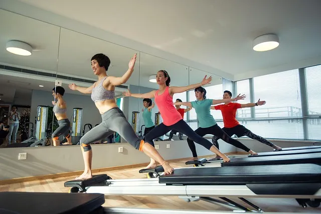
Yoga has consistently shown improvements in lung function and breathing mechanics in studies that included spirometry and respiratory muscle testing. Pranayama practices train extended breath cycles and diaphragmatic engagement. Asanas like cobra, sphinx, cat-cow, and warrior series all mobilize the thoracic spine and ribcage in ways that directly improve respiratory mechanics. A meta-analysis of yoga in healthy adults found significant improvements in FVC, FEV1, and respiratory muscle endurance from 8-12 week programs.
Pilates emphasizes the connection between core stability, spinal alignment, and breathing — which matches the anatomical reality described throughout this article. The Pilates breathing cue to inhale laterally (into the sides of the ribcage) specifically targets the posterior and lateral expansion pattern associated with optimal diaphragmatic breathing. Research in COPD patients found Pilates-based breathing training improved respiratory mechanics more than standard diaphragmatic breathing training alone.
The Feldenkrais Method and Alexander Technique both directly address habitual postural and breathing patterns through awareness-based movement education. Multiple case studies and some controlled trials have shown improvements in breathing pattern and respiratory function following these approaches — likely through interruption of the chronic neuromuscular habits that maintain dysfunctional posture and breathing.
Exercise Prescription for Postural Breathing Optimization
- Daily: Thoracic foam rolling (2-3 minutes at mid-back and upper back). The single most impactful acute intervention for thoracic mobility.
- Daily: Chin tucks (3 sets of 10 repetitions with 5-second holds). Restores cervical neutral alignment and activates deep cervical flexors.
- Daily: Crocodile breathing (5 minutes prone diaphragmatic breathing). Establishes posterior expansion pattern.
- 3x/week: Thoracic rotation stretches (10 reps each direction). Maintains rotational mobility essential for ribcage breathing.
- 3x/week: W exercise or prone Y-T-W (3 sets of 10 reps). Builds thoracic extensor strength to counteract kyphosis.
- 2x/week: Doorway pectoral stretch (3 holds of 60 seconds). Reduces anterior chain restriction limiting ribcage expansion.
Anatomy Breathing Posture Q&A
How long does it take to correct forward head posture? Meaningful improvement in head position and breathing mechanics is typically measurable at 6-8 weeks of consistent daily practice. Complete correction — if “correction” is even the right word, given that moderate deviations can still be functional — takes months to years of habit change. The relevant timeframe isn’t weeks. It’s the rest of a person’s life. Sustainable postural habits, not temporary exercise programs, are what matter long-term.
Does sitting at a desk inevitably cause breathing problems? Prolonged static sitting creates cumulative postural loading, but the research suggests frequent position changes — standing, stretching, walking — can significantly mitigate these effects even without changing the total sitting time. The ideal isn’t to sit less but to sit differently: varied positions, frequent breaks (every 20-30 minutes), ergonomic setup that supports neutral spine, and deliberate postural reset exercises during the day.
Can poor breathing posture cause back pain? Yes, through multiple mechanisms. Altered rib mechanics change thoracic and lumbar loading patterns. Diaphragm dysfunction reduces intra-abdominal pressure that supports the spine. Upper chest breathing increases paraspinal muscle tension. Studies have found chronic low back pain patients have measurably reduced diaphragm excursion and altered respiratory muscle coordination compared to pain-free controls — and breathing retraining improves back pain outcomes in this population.
Is there a relationship between posture, breathing, and anxiety? Yes, and the causality runs in both directions. Anxious breathing patterns (shallow, rapid, upper chest) activate sympathetic responses that increase anxiety. And anxious states create postural patterns (shoulders up and forward, chest collapsed) that further restrict breathing. Correcting postural breathing is a physiological intervention for anxiety — one the research supports alongside behavioral approaches.
What about breathing during weight training? The Valsalva maneuver — deep breath, breath hold during maximal effort, exhale at the sticking point — uses the diaphragm to create intra-abdominal pressure that supports the spine during heavy lifting. Appropriate for near-maximal loads. For moderate-intensity training, the standard cue is exhale during the effort phase, inhale during the recovery phase. Maintaining 360-degree ribcage expansion during all training positions (thoracic volume held even when flexed or rotated) protects spinal integrity.
Are commercial posture correctors helpful? The evidence is mixed. Passive bracing — wearing a harness that prevents forward shoulder position — may reduce short-term muscle fatigue and pain but doesn’t develop the active motor control needed for lasting postural change. In some studies, passive bracing has been associated with weakness of the postural muscles it unloads. Active approaches — exercises building the strength and motor patterns for self-maintained posture — are consistently more effective long-term.
How does posture affect voice and speech breathing? Significantly. The resonance and projection of the voice depend on breath support — the controlled exhalation that carries sound. Thoracic kyphosis and forward head posture reduce the efficiency of breath support by compromising the breathing foundation. Singers, actors, and public speakers consistently show better postural alignment than the general population — not incidentally, but because their training demands effective breathing mechanics, and postural alignment is the structural prerequisite.
The Seated Work Position and Breathing Optimization

Seat height and hip angle are critical. When hips sit at 90 degrees (standard chair height) or below, the pelvis tends to tilt posteriorly, flattening lumbar lordosis and compressing the lower thoracic region. Raising the seat or using a wedge cushion to create a 100-110 degree hip angle lets the pelvis sit at neutral tilt, preserving lumbar lordosis, and creating the spinal foundation for diaphragmatic breathing. This single adjustment can meaningfully improve breathing mechanics for people who spend most of the day seated.
Screen height affects head and thoracic position. A screen that requires the head to tilt downward (common with laptops) drives forward head posture and thoracic flexion. Raising the monitor so the top of the screen sits at or slightly below eye level dramatically reduces the tendency to flex forward. Laptop stands combined with external keyboards are the most cost-effective fix — they convert a posture-impairing device into one usable in optimal position.
Active seating — sit-stand desks, balance boards, active seating chairs, or even a simple practice of frequent position changes — maintains thoracic mobility and prevents the static loading that progressively compresses ribcage mechanics. Research on standing desks has found improvements in spinal posture markers and reduced musculoskeletal discomfort, though the breathing-specific benefits haven’t been directly studied in large trials. The logic is straightforward: standing eliminates the seated hip-flexion-driven posterior pelvic tilt and gives the thoracic spine more freedom of movement.
The two-minute postural reset — standing, rolling shoulders back and down, performing chin tuck, taking five slow diaphragmatic breaths — takes almost no time and provides a measurable reset of the postural pattern after extended seated work. Setting a timer to perform this reset every 30-45 minutes interrupts the progressive accumulation of flexion loading that would otherwise compound across the entire workday.
Breathing Posture Across the Lifespan
The trajectory of breathing posture changes across life in predictable ways that create specific intervention windows. In childhood, breathing is typically diaphragmatic and effortless — the natural pattern before years of sitting, screen use, and stress-driven accessory muscle breathing create the dysfunctions described throughout this article. Intervening in adolescence — before these patterns fully set — has the highest return per unit of intervention effort.
In adults aged 20-50, the primary drivers of postural breathing dysfunction are occupational (prolonged sitting) and stress-related (chronic sympathetic activation driving upper chest breathing). This is the window where intentional habit change — the exercise protocols and ergonomic modifications described above — can prevent the progressive structural changes that make correction harder in later decades.
In adults over 60, structural changes — thoracic kyphosis often driven by vertebral compression fractures from osteoporosis, reduced thoracic soft tissue flexibility from cumulative shortening, decreased diaphragm strength from aging and deconditioning — make the respiratory mechanical compromise more significant and the correction more challenging, though still relevant. Studies have found thoracic mobility training in older adults produces significant improvements in respiratory function and quality of life, suggesting the system retains plasticity even in later decades.
The message is consistent across the lifespan: postural breathing dysfunction is not a fixed state determined by age and history. It’s a dynamic variable that responds to appropriate loading — mobility work, strengthening, breathing pattern retraining, ergonomic modification. The earlier the intervention, the more potential for prevention rather than correction. But correction is meaningful and achievable at any age. The anatomy that creates the problem is the same anatomy that responds to the solution — it just requires the consistent application of the right inputs.
The intersection of posture and breathing is where structural mechanics meets functional physiology. Optimizing this intersection — through the specific interventions described throughout this article — isn’t about aesthetics or athletic performance optimization. It’s about the quality of every breath, for the rest of a person’s life. That’s 23,000 daily opportunities to do better, or to keep accumulating dysfunction. The choice, repeated daily, is literally structural.
The Neuroscience of Breathing and Posture
The connection between posture and breathing extends into the nervous system in ways that go beyond simple mechanics. The vagus nerve — the primary conduit of parasympathetic signaling from the brain to the body — passes through the thoracic inlet and is affected by the position of the cervical spine and thoracic structures. Chronic forward head posture and thoracic compression can reduce vagal tone — cutting into the parasympathetic capacity that governs rest-and-digest function, heart rate variability, and airway regulation.
The right vagus nerve branches to the right lung and the left to the left lung — these branches regulate bronchomotor tone (airway caliber) and mucociliary clearance. Optimal vagal function supports airway dilation and normal mucus clearance. Reduced vagal tone from postural compression or chronic sympathetic dominance contributes to increased bronchomotor tone and potentially reduced airway clearance efficiency. One mechanism by which the breathing retraining and postural correction described in this article produce respiratory benefits beyond the purely mechanical.
Proprioceptive input from thoracic joint mechanoreceptors influences the central respiratory pattern generator in the brainstem — the neural circuit determining baseline breathing rate and depth. Restricted thoracic joint mobility reduces the proprioceptive input from this region, potentially contributing to the shallow, rapid breathing patterns common in people with significant thoracic restriction. Manual therapy that restores thoracic joint mobility can produce acute improvements in breathing pattern — a finding replicated across multiple studies of spinal manipulation and mobilization in patients with breathing dysfunction.
The phrenic nerve — the motor nerve to the diaphragm — originates from cervical levels C3-C5. Chronic upper cervical compression from forward head posture can create subtle phrenic nerve irritation that manifests as diaphragm dysfunction. A less common but documented mechanism of breathing impairment in people with significant cervical dysfunction, and it explains why cervical spine treatment sometimes produces unexpected improvements in breathing quality.
Motor cortex mapping for breathing — the cortical representation of respiratory muscles — shows voluntary control of breathing mechanics (diaphragmatic breathing practice, deliberate three-dimensional expansion, conscious breath holds) can expand and strengthen the cortical representation of respiratory muscles. The same neuroplasticity principle that governs skill learning in other motor domains. Deliberate practice of optimal breathing mechanics — done consistently over weeks and months — literally changes the brain’s representation of breathing, making the new pattern more automatic and the old pattern less default. Which is why breathing retraining produces lasting changes: it rewrites the motor program.
Understanding this neuroscience matters because it explains why passive postural correction (braces, pillows, furniture adjustments alone) produces limited lasting results while active motor retraining produces durable change. Changing the structural input (ergonomics, foam rolling) creates the precondition. Retraining the motor program (breathing exercises, postural exercise, practice of new patterns under load) is what establishes the new default. Both are necessary. Neither alone is sufficient.
Putting It All Together: The Integrated Breathing-Posture Protocol
The interventions described throughout this article are most effective as an integrated protocol rather than isolated techniques applied without structure. Here’s a daily framework addressing all the key elements without requiring extraordinary time investment.
Morning (10 minutes): Begin with two minutes of thoracic foam rolling at mid and upper back. Follow with chin tucks (3 sets of 10) and doorway pectoral stretch (2 holds per side, 30 seconds). Complete with five minutes of crocodile breathing prone — diaphragmatic expansion directed downward and laterally. This sequence restores overnight postural restriction and establishes the breathing pattern for the day before the habits of desk work set in.
Workday (2 minutes every 30-45 minutes): The postural reset — stand, roll shoulders back and down, perform 5 chin tucks, take 5 diaphragmatic nasal breaths with extended exhale (4 counts in, 6 counts out). This interrupts the progressive accumulation of postural flexion loading and maintains the breathing pattern established in the morning routine. Frequency matters more than the duration of each reset.
Evening (5-10 minutes): Thoracic rotation stretches (10 reps each direction) and cat-cow exercises (10 cycles), addressing the rotational and flexion-extension mobility that daily sitting restricts. Add W exercise or prone Y-T-W strengthening (3 sets of 10) for thoracic extensor strength three times per week. End with 4-7-8 breathing (4 cycles) to complete the transition from sympathetic daytime mode to parasympathetic rest-and-recovery mode.
Total daily investment: 20-25 minutes, distributed throughout the day. The cumulative effect on breathing mechanics, spinal mobility, and postural pattern over months is substantial — routinely reported by people who maintain this protocol as one of the most impactful changes they’ve made to how they feel day to day. Not because any individual session is dramatic, but because the compound effect of addressing breathing mechanics in 23,000 daily breaths, consistently, over years, accumulates into a fundamentally different physical baseline.
The breath is always available as a tool for recalibration, for structural maintenance, for physiological signaling. The posture brought to each breath determines how effectively that tool can be used. Get the structure right, and the function follows. Not a metaphor. Anatomy, applied consistently, over a lifetime.
Research from the field of exercise physiology has consistently found that breathing mechanics account for a surprisingly large portion of exercise-related fatigue at submaximal intensities. People who breathe more efficiently — deeper, slower, more diaphragmatically — use less energy for the act of breathing itself, leaving more available for activity. In fit adults, the respiratory muscles consume approximately 1% of cardiac output at rest and up to 10% during maximal exercise. In deconditioned individuals with dysfunctional breathing patterns, the respiratory fraction runs higher at any given workload. Improving breathing mechanics doesn’t just feel better. It measurably frees up metabolic resources for everything else the body needs to do.
The practical implications extend beyond exercise. The daily cognitive and metabolic cost of suboptimal breathing accumulates into the background fatigue many people accept as normal aging. Someone who’s spent decades breathing with accessory muscles, hyperventilating subtly, and sleeping in positions that compromise the airway is genuinely more fatigued than they need to be. Correcting the breathing-posture relationship is one of the few interventions that reliably produces a sense of increased energy patients often describe as feeling years younger. Not because it reverses aging, but because it removes a continuous drain on metabolic resources that shouldn’t have been there in the first place.
The actionable finding is that the body is responsive. The muscles that control posture and breathing are highly adaptable. The neural patterns that maintain dysfunctional defaults are rewritable. The connective tissue that restricts thoracic mobility responds to consistent stretching. None of this is instant, and none of it is passive — it requires the daily practice described in this article, applied consistently enough for the new patterns to become the new default. That transition takes roughly 8-12 weeks to produce measurable structural changes, and months to years to fully consolidate. But it begins with the next breath — taken with more awareness, more depth, and more structural support than the one before it.
The clinical evidence base for postural breathing interventions continues to grow as researchers recognize that breathing dysfunction is both a cause and a consequence of musculoskeletal and systemic conditions. Physical therapists working in pulmonary rehabilitation increasingly incorporate postural assessment and thoracic mobility training alongside traditional breathing exercises. Occupational therapists evaluate workstation ergonomics for breathing implications. Sports physiologists assess breathing mechanics in athletes as a performance variable. This integration — treating the breathing system as mechanically embedded in the structural body rather than as an isolated respiratory function — reflects the anatomical reality that has always been true and is only now being fully operationalized in clinical practice.
The practical takeaway: the way a body is held determines the quality of every breath it takes. The quality of every breath determines the efficiency of oxygen delivery, autonomic tone, physiological recovery capacity, and long-term respiratory health. Improving breathing posture is not a niche wellness pursuit. It’s basic physiological maintenance for anyone who intends to function at a high level for a long time. The tools are accessible, the evidence is solid, and the returns compound indefinitely. Start today, with the breath being taken right now.
The relationship between breathing and posture is bidirectional in another important sense: improving breathing habits improves posture. When breathing happens diaphragmatically and fully, the resulting pressure dynamics in the thoracic and abdominal cavities create an internal postural support system that reduces the load on passive structures like ligaments and intervertebral discs. The diaphragm, pelvic floor, deep abdominals, and multifidus muscles work as an integrated pressure management system — the dynamic neutral spine support that no external brace can replicate. Training proper breathing is, simultaneously, training the core stability system. These aren’t separate interventions. They’re the same intervention, approached from different angles of the same anatomy. Mastering diaphragmatic breathing is also a significant investment in spinal health, postural endurance, and the basic structural foundation that keeps a person upright and functional for decades longer than someone who neglects both.
The Practical Framework: Applying Anatomy Breathing Posture In Real Life
References
Editorial StandardsCorrectionsMedical DisclaimerAbout Our ContentAffiliate DisclosureSite Map